Management of Palatally Positioned Impacted Mesiodens: 2 Case Reports
Sachin A Gunda, Anand L Shigli, Anil T Patil, Bhargav S Sadawarte, Ankita R Hingmire and Priyanka A Jare
DOI10.21767/2469-2980.100038
Sachin A Gunda, Anand L Shigli, Anil T Patil*, Bhargav S Sadawarte, Ankita R Hingmire and Priyanka A Jare
Department of Pedodontics and Preventive Dentistry, Bharati Vidyapeeth Deemed University Medical College and Hospital, Sangli, India.
- *Corresponding Author:
- Anil T Patil
Associate Professor, Department of Pedodontics and Preventive Dentistry
Bharati Vidyapeeth Deemed University Dental College and Hospital
Sangli, India-416414.
Tel: +91 9850983500
E-mail: dranilp0888@gmail.com
Received date: January 05, 2017; Accepted date: March 08, 2017; Published date: March 13, 2017
Citation: Sachin AG, Anand LS, Anil TP, et al. Management of Palatally Positioned Impacted Mesiodens: 2 Case Reports. J Orthod Endod. 2017, 3:1. doi: 10.21767/2469-2980.100038
Copyright: © 2017 Anil TP, et al. This is an open-access article distributed under the terms of the Creative Commons Attribution License, which permits unrestricted use, distribution, and reproduction in any medium, provided the original author and source are credited.
Abstract
By definition the extra teeth present in the oral cavity are the supernumerary teeth which are found in any area of the oral cavity. Sometimes Supernumerary teeth are present even in the anterior region of the maxilla which is called as mesiodens. Mesiodens are the most common supernumerary teeth, occurring in 0.15–1.9% of the population. The exact etiology of mesiodens is not fully understood, but proliferation of the dental lamina and genetic factors can be the reason. Mesiodens can be either paired or single, unerupted or impacted which can be unaesthetic and most of the time associated with complications like midline diastema, rotation, displacement, and root resorption and cyst formation. It should be properly diagnosed for the appropriate management to reduce the complications to the developing dentition. In the present cases mesiodentes were found in the anterior maxilla, one erupted and another unerupted and inverted in one case and unerupted causing resorption of root of permanent central incisor in another case. In these cases mesiodentes are prophylactically extracted to prevent its adverse effects on developing dentition.
Keywords
Discoloration; Impacted mesiodens; Mesiodens; Supernumerary teeth
Introduction
The extra teeth present in addition to the normal dentition are the supernumerary teeth. This state is called as Hyperdontia. The existence of first report of supernumerary tooth has been found around 23–79 AD. The commonest form of supernumerary teeth frequently found is the mesiodens [1]. In 1917, Balk gave the term “mesiodens” which indicated a supernumerary tooth present mesial to both central incisors and also appeared as peg shaped with either inverted or normal position. Two types of mesiodens are found depending on their shape and size. Eumorphic tooth is the first type which is nothing but a regular morphology and similar to the central incisor. Dysmorphic teeth being the second type which has dissimilar shapes and sizes and are further classified into supplemental, odontomas, conical, and tuberculate [2].
In the literature, the incidence of supernumerary teeth vary from 0.1-3.6% in permanent dentition [3,4]. Prevalence is lower in deciduous teeth amounting to 0.3–0.8% [5,6]. Males are affected more frequently, with varying ratios from 2:1 and 6:1. A constant gender distribution appears in the deciduous dentition.
The etiology behind the development of these supernumerary teeth is not fully understood. Various etiological theories have been assumed for the development of supernumerary teeth. One theory suggests, organized and an excessive growth of dental lamina to form a third tooth germ splitting of the permanent tooth bud [7-10]. Dichotomy of the tooth bud is the reason for supernumerary teeth as suggested by another theory [11]. One more theory, well supported in the literature, is the hyperactivity theory, suggesting that the hyperactivity of dental lamina is the reason for supernumerary teeth formation [12]. Genetics are also instrumental for the occurrence of supernumerary teeth, as these anomalies are more common in the family of affected children than in the general population [13]. Few syndromes like Hallermann-Streiff's syndrome, Gardner's syndrome, Cleidocranial dysplasia, cleft lip and palate, oro-facial digital syndromes, etc. usually have supernumerary teeth present [8].
Mesiodens, which are usually unaesthetic frequently, causes either malposition or delayed eruption of adjacent central incisors, midline diastema, caries, and odontogenic cysts, gingival and periodontal problems. They can be the reason for impaction, resorption, and displacement of central incisors and may extend into the nasal cavity. Hence, early diagnosis and extraction of mesiodens is very essential to avoid these types of complications.
Clinical Findings and Case Report
Case 1
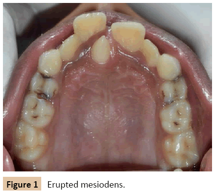
A 10-year-old patient reported to the department of Pedodontics, with a complaint of forwardly placed upper front teeth with an extra tooth in between them. After thorough examination, it was found that medical and family histories were noncontributory. Extra oral examination didn't reveal any abnormalities. Intra oral examination revealed mixed dentition. Patient had class 1 molar relation on both side with very mild upper anterior crowding. A Mesiodens (Figure 1) was seen palatal to maxillary left central incisor; it was conical in shape (mesiodens) measuring about 5 mm mesio-distally.
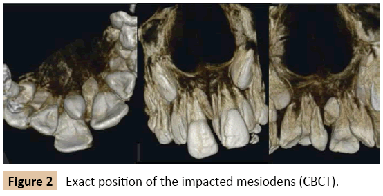
Occlusal radiograph was taken to rule out the possibility of multiple supernumerary teeth and we found out another unerupted and impacted inverted mesiodens, mesial to the maxillary right central incisor. After obtaining intra- oral radiograph of maxillary right and left central incisors, we observed that the impacted and inverted mesiodens was in relation to maxillary right Central Incisor. Patient was advised to undergo Cone Beam Computed Tomography (CBCT) in order to obtain the exact position of the impacted mesiodens (Figure 2).
There were two mesiodens, one erupted and another impacted and inverted. In this case, patient's age was 10 years and adjacent permanent central incisors were totally erupted, so the treatment plan was to extract both erupted and unerupted mesiodentes under local anaesthesia. Both informed and written consent were taken from the parent's prior to surgical procedure.
1) Intra-alveolar extraction (forceps technique) of erupted supernumerary teeth was done.
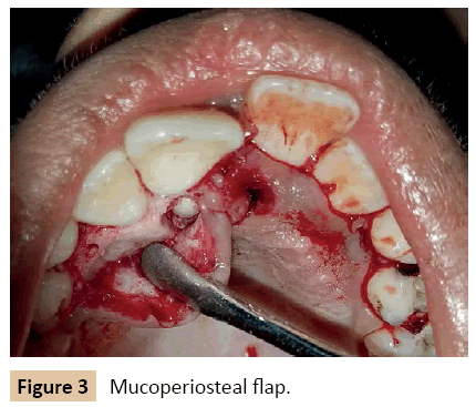
2) Then surgical extraction of the unerupted mesiodens was carried out by raising mucoperiosteal flap: first intracrevicular incision was made from 1st premolar to contralateral 1st premolar on palatal side then a mucoperiosteal flap was raised (Figure 3).
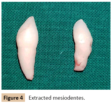
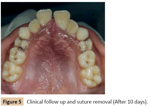
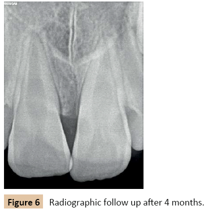
Adequate amount of bone was removed using rotary cutting instruments after elevation of the flap. The mesiodens was removed (Figure 4) and the extraction socket was checked for any pathological tissue. The flap was relocated and interrupted sutures were placed for a week. After 10 days sutures were removed and patient was recalled after four months for followup (Figures 5 and 6).
Case 2
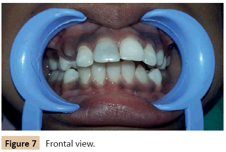
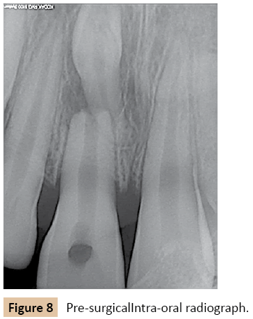
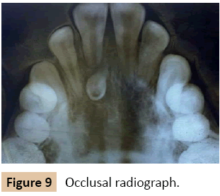
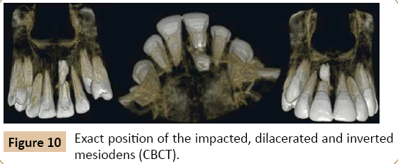
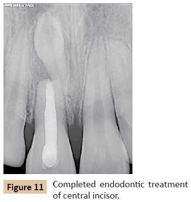
A 10-year-old male patient reported to the dept. of Pedodontics and preventive dentistry, with a complaint of forwardly placed upper front teeth along with discolored right front tooth (Figure 7). He did not have any history of trauma to upper anterior region. There were no any signs or symptoms related to trauma from occlusion. After thorough clinical examination, the periapical radiograph was taken (Figure 8), which exposed root resorption of the right central incisor. There was an impacted mesiodens approaching the root of right central incisor. Patient had been advised for occlusal radiograph and CBCT to know the exact position of the impacted, dilacerated and inverted mesiodens (Figures 9 and 10). Endodontic treatment of the right central incisor was completed (Figure 11).
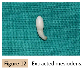
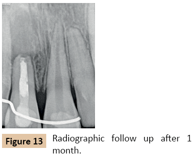
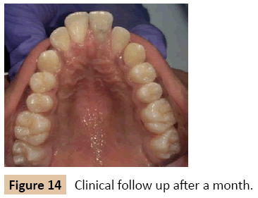
Surgical extraction was considered as a treatment. After giving local anesthesia, the palatal flap was raised from distal aspect of first premolar on left side to distal aspect of first premolar on right side. Extraction of the mesiodens was carried out after locating the crown, with minimal bone cutting (Figure 12), and sutures were placed. Splinting was carried out in order to stabilize right Central Incisor as tooth showed grade I mobility. Excellent healing was observed after two weeks. Patient came for the follow up after 1, 3 and 5 months (Figures 13 and 14).
Discussion
Supernumerary tooth is an extra tooth present in any area of the oral cavity which can be found both in deciduous or permanent dentition. The most commonly found supernumerary teeth are the mesiodens, maxillary distomolars, maxillary paramolars, mandibular parapremolars, mandibular para and disto molars, and maxillary parapremolars [14,15]. Most of the time, impacted supernumerary teeth remain clinically silent and are diagnosed during radiographic examination. They can cause complications and require an immediate treatment.
Numerous theories have been proposed about the aetiology of supernumerary teeth which include phylogenetic, hyperactivity of dental lamina, and dichotomy of tooth bud. Nevertheless, the precise reason of development of these teeth is still not known and not completely recognized. According to “Phylogenetic theory”, the origin and development of supernumerary teeth is due to atavism of extinct ancestral tissues. The tooth germ dichotomy theory said that the dental lamina sometimes divides into two or more parts, thus leading to the development of an extra tooth which is either similar or sometimes different in size. As said by “hyperactivity of dental lamina” theory, the formation of supernumerary tooth/teeth is due to over-proliferation of remnants of dental lamina. As these supernumerary teeth are seen commonly among the relatives of affected individuals when compared to the general population, heredity is one of the etiology [2,15].
A mesiodens is a supernumerary tooth which erupts in between the maxillary central incisors and is frequently positioned palatally. Only some of them present within the arch or labially. 80% of impacted mesiodens are found palatally, 6% are located labially and 14% are positioned between the roots of the permanent central incisors [16]. The clinical complications of mesiodens include delayed eruption of permanent incisors, axial rotation or inclination of adjacent teeth or mesiodens, midline diastema, intraoral infection, root anomaly, and pulpitis of the mesiodens [17-20]. A cystic alteration is observed in 4–9% of the supernumerary cases, with the anterior maxilla being affected in 90% cases [21,22]. This indicates surgical removal of the supernumerary teeth [23].
Treatment of mesiodens varies on several factors whether to manage surgically or to observe the condition. The major factor is the child's age, in the very young patient, the ability to stand a surgical procedure is of chief concern. The advantage of early management is essential for the long-term effect to avoid unpleasant experience that may have psychological effect on patient [24]. Second is the closeness of the mesiodens to the incisors and stage of development of the dental structures adjoining teeth? In cases of undeveloped root, one should know the danger of surgical trauma to the developing roots of the permanent incisors and the possible damage to the future dental growth. Mesiodens positioned closely to the developing permanent incisors may change the location of the succedaneous tooth bud, hamper eruption, and/or change root development; whereas, surgical extraction of the same mesiodens may cause the same sequel with surgical trauma. In cases where the surgical approach endangers the sustainability of sensitive developing tooth bud, it may be suitable to hold-up the treatment. At last, the dentist must assess the relative position of the mesiodens within the jaw whether mesiodens is labially or palatally placed.
In surgical procedure, access to the mesiodens must be considered corresponding to the quantity of bone amputation and possible damage to existing incisors. In children, eruption of mesiodens is possible and complete eruption is occasional, some mesiodens may erupt incompletely and hence more favorable surgical approach may be accomplished with time [24,25].
In the first case, one erupted and unerupted mesiodentes were present. No signs and symptoms were present associated with an unerupted mesiodens. On the contrary, root resorption was significant with respect to right central incisor in another case. When no clinical sign is evident and the mesiodens is asymptomatic and impacted, it can cause many pathological findings like granuloma or cyst as already discussed, and the young children are more prone to these manifestations. This is because the permanent dentition is not developed yet at this age and occlusion is not established, chances of pathologies in young children is highly associated with unerupted supernumerary teeth because it may act as nidus for cystic transformation or spread of infection. Hence prophylactic extraction of these mesiodentes is indicated in order to avoid problems and minimize the complications. In the first case the timing for surgical extraction of both mesiodentes was suitable since both maxillary central incisors were totally erupted showing complete root formation.
In the second case, mesiodens was associated with pathological root resorption of the right maxillary permanent central incisor leading to non-vitality and successive discoloration of the central incisor. Mesiodens was impacted palatally and was removed to prevent any further harm to the central incisor or cyst formation. Endodontic treatment of central incisor was done. The patient was gratified with the treatment.
Conclusion
Supernumerary teeth are of huge concern to both dentist and patient because of its potential problems and complications. Radiographic evaluation of erupted supernumerary teeth is important in the accidental detection of unerupted mesiodens. On diagnosis, every case should be treated properly to lessen problems to the developing tooth buds and dentition. Careful history taking, clinical and radiographic examinations can provide important information required for the diagnosis of such conditions. Once surgical removal of mesiodens is advised, long-term follow-up of treated case is required.
References
- Sheikh Z, Manzoor A, Amir N (2014) Mesiodens: A common supernumerary tooth: Report of management of a case with two mesiodens. Int Dent J Stud Reason2:41-46.
- Russell KA, Folwarczna MA (2003) Mesiodens-Diagnosis and management of a common supernumerary tooth. J Can Dent Assoc 69:362-366.
- Brook AH (1974) Dental anomalies of number, form, and size: Their prevalencein British Schoolchildren. J IntAssoc Dent Child5:37-53.
- Davis PJ (1987)Hypodontia and hyperdontia of permanent teeth in Hong Kong school children. Commun. Dent Oral Epidemiol15:218-220.
- Brabant H (1967) Comparison of the characteristics and anomalies of the deciduous and the permanent dentitions. J Dent Res46:897-902.
- Ravn JJ (1971) Aplasia, supernumerary teeth and fused teeth in the primary dentition. An epidemiologic study. Scand J Dent Res79:1-6.
- Ibricevic H, Al-Mesad S, Mustagrudic D, Al-Zohejry N (2003) Supernumerary teeth causing impaction of permanent maxillary incisors: consideration of treatment. J ClinPediatr Dent 27:327-332.
- Zhu JF, Marcushamer M, King DL, Henry RJ (1996) Supernumerary and congenitally absent teeth: A literature review. J ClinPediatrDent 20:87-95.
- Taylor GS (1972) Characteristics of supernumerary teeth in the primary and permanent dentition. Dent Pract Dent Rec 22:203-208.
- Luten JR (1967)The prevalence of supernumerary teeth in primary and mixed dentitions. J Dent Child34:346-353.
- Liu JF (1995) Characteristics of premaxillary supernumerary teeth: A survey of112 cases. ASDC J Dent Child 62:262-265.
- Levine N (1961)The clinical management of Supernumerary teeth. J Can DentAssoc28:297-303.
- Rosenzweig KA, Garbarski D (1965) Numerical aberrations in the permanent teeth of grade school children in Jerusalem. Am J PhysAnthropol23:277-283.
- Kokten G, Balcioglu H, Buyukertan M (2003) Supernumerary fourth and fifth molars: A report of two cases. J Contemp Dent Pract4:67-76.
- GaphorSM, Abdulkareem SA, Abdullah MJ (2014)Unilateral maxillary distomolar: A case report and review of the literature. J Dent Med Sci 13:17-20.
- Khandelwal P, Hajira N (2015) Mystery behind discoloration of central incisor: An inverted, labially impacted mesiodens. Int J Oral Health Sci 5:129-132.
- Gallas MM, García A (2000) Retention of permanent incisors by mesiodens: a family sffsir. Br Dent J 188: 63-64.
- Kim SG, Lee SH (2003) Mesiodens: a clinical and radiographic study. J Dent Child 70: 58-60.
- Tyrologou S, Koch G, Kurol J (2005) Location, complications and treatment of mesiodentes-a retrospective study in children. Swed Dent J 29: 1-9.
- Von Arx T (1992) Anterior maxillary supernumenary teeth: a clinical and radiographic study. Aust Dent J 37: 189-195.
- Primosch RE (1981) Anterior supernumerary teeth-assessment and surgical intervention in children. Pediatr Dent 3: 204-215.
- Lustmann J, Bodner L (1988)Dentigerous cysts associated with supernumerary teeth. Int J Oral MaxillofacSurg17:100-102.
- Mitchell L, Bennett TG (1992) Supernumerary teeth causing delayed eruption: a retrospective study. Br J Orthod 19: 41-46.
- Henry RJ, Post AC (1989)A labially positioned mesiodens: Case report. Pediatr Dent 11:59-63.
- Sandhyarani B, Huddar D (2012) Mystery behind malocclusion: Report of two mesiodens cases. J Dent Med Sci 2:46-49.
Open Access Journals
- Aquaculture & Veterinary Science
- Chemistry & Chemical Sciences
- Clinical Sciences
- Engineering
- General Science
- Genetics & Molecular Biology
- Health Care & Nursing
- Immunology & Microbiology
- Materials Science
- Mathematics & Physics
- Medical Sciences
- Neurology & Psychiatry
- Oncology & Cancer Science
- Pharmaceutical Sciences
