Pursuit for Optimum Skeletal Expansion: Case Reports on Miniscrew Assisted Rapid Palatal Expansion (MARPE)
Neeraj Eknath Kolge, Vivek J Patni, Sheetal S Potnis, Swapnagandha Ravindra Kate, Floyd Stanley Fernandes and Chetna Dadarao Sirsat
DOI10.21767/2469-2980.100059
Neeraj Eknath Kolge*, Vivek J Patni, Sheetal S Potnis, Swapnagandha Ravindra Kate, Floyd Stanley Fernandes and Chetna Dadarao Sirsat
M.G.M. Dental College & Hospital, Navi Mumbai, Maharashtra, India
- *Corresponding Author:
- Dr. Neeraj Eknath Kolge
M.G.M. Dental College & Hospital
Navi Mumbai, Maharashtra, India
E-mail: neerajkolge11@gmail.com
Received Date: June 07, 2018; Accepted Date: June 29, 2018; Published Date: July 05, 2018
Citation: Kolge NE, Patni VJ, Potnis SS, Kate SR, Fernandes FS, et al. (2018) Pursuit for Optimum Skeletal Expansion: Case Reports on Miniscrew Assisted Rapid Palatal Expansion (MARPE). J Orthod Endod 4:9. DOI: 10.21767/2469-2980.100059
Abstract
Incorporation of mini screws in a conventional RPE appliance transforms it into a MARPE appliance. Mini screws ensure maximum skeletal expansion, keeping the dental expansion and resultant side effects to a minimum. Various designs have been recommended by authors around the globe; exclusively bone borne, teeth-bone borne and tissue-bone borne with two/ four mini screws in the assembly. Paramedian area 3 mm lateral to the suture in 1st premolar region is considered the most appropriate site for placement of mini screws. Anterior screws are placed in the rugae area while posterior screws in the para-midsagittal area. This article (case series) describes three cases treated with MARPE appliance designs and protocols.
Keywords
MARPE; Rapid palatal expansion; Maxillary skeletal expansion
Introduction
Incorporation of mini screws in a conventional rapid palatal expansion (RPE) appliance transforms it into a miniscrew assisted rapid palatal expansion (MARPE) appliance [1]. Mini screws ensure maximum skeletal expansion, minimizing the dental side effects [2]. Various designs have been recommended by many authors [3,4] without any dental support (exclusively bone borne), with support from teeth (teeth-bone borne) and two/ four mini screws.
Stress distribution trajectories are mainly along three buttresses in the maxilla; namely zygomaticomaxillary, nasomaxillary and pterygomaxillary. Major disadvantages of conventional RPE appliances include tipping of anchor teeth [5], limited skeletal movement [6], undesirable tooth movement [7], root resorption [8], bony dehiscences and fenestrations as well as post expansion relapse [9].
As, conventional RPE appliances transmit the expansion forces through the teeth, alveolar bone bending and tipping of buccal segments is unavoidable. This not only takes up significant activation of the appliance, but also reduces the true skeletal effect as well as creates clockwise rotation of mandible, thus opening the bite.
Thus, MARPE appliance is beneficial in adult patients with more sutural resistance to skeletal expansion and even in young patients by preventing/ minimizing dental tipping thus avoiding further increase in the vertical dimension and other aforementioned side effects.
Location of Mini Screws
Factors like convenient access, low risk of damage to the surrounding anatomical structures [10-12], high quality cortical bone and thin mucosa confirming adequate stability [13,14]. makes paramedian area (3 mm lateral to the suture in 1st premolar region) the most appropriate site for placement of mini screws [15-17].
Mini Screw Configuration
Dimension of two/ four mini screws as per the design were selected as follows
Anterior screws (Rugae area): 1.8 x 10 mm (FavAnchorTM SAS, India)
Posterior screws (Para-midsagittal area): 1.8 x 8 mm (FavAnchorTM SAS, India)
The lengths were chosen considering height of insertion slot, space between the appliance and the palate, thickness of the palatal mucosa and desired 5-7 mm of bone engagement. Intention was to help bicortical engagement [18], thus aiding for better stability of the mini screws. TAD placement with a conventional straight driver or an engine mounted driver problematic sometimes for the reasons of directional control and lack of torque to drive the implant in hard palatal bone.
A dedicated palatal driver [(L’il One, FavAnchorTM SAS, India)] is used to maintain adequate insertion angulation and torque while placing the mini screws. This unique design of the driver makes it very convenient to place the palatal implants with great ease and precision.
Fabrication
The size of expansion screw was selected taking into consideration it’s close adaptation to the palatal vault. MARPE expander was fabricated by soldering rigid connector wires on the expansion screw. Accurate and passive adaptation of the insertion slot on the palate ensured perpendicular positioning of mini screws. Posterior implant, in the 4 screw design, should be placed as close to the body of the screw, as bone thickness reduces significantly as we go more posterior on the palate.
Activation Protocol/ Schedule
Activation was initially done for 2 turns/ day (360° x 2) till development of diastema, followed by 1 turn/ day till sufficient expansion has been achieved [3].
Activation schedule was followed as described (Table 1) [4].
| Age of the patient | Initial expansion rate | Expansion rate after opening of the diastema |
|---|---|---|
| Early teens | 3 turns/ week | 3 turns/ week |
| Late teens | 1 turn/ day | 1 turn/ day |
| Adults | 2 turns/ day | 1 turn/ day |
| Older patients (>30 years) | >2 turns/ day | 1 turn/ day |
Table 1: Activation schedule.
Treatment Objectives
The treatment objectives were to as follows:
1. To correct transverse maxillary deficiency
2. To maximizing skeletal expansion
3. To minimizing buccal tipping
4. To establish acceptable buccal occlusion and
5. To maintain sound periodontal and bone support
Case 1
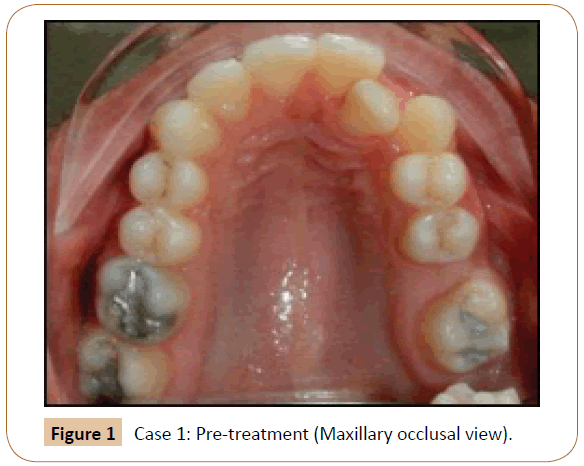
17 year 4 months old, female patient presented with moderately crowded upper and lower anteriors. Clinical examination revealed instanding maxillary and mandibular left lateral incisors, missing permanent maxillary first molar and dark buccal corridors. She presented with a skeletal Class II pattern and a clockwise rotational tendency (Figure 1).
Appliance design
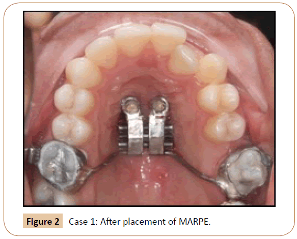
Two mini screws (1.8 x 10 mm, FavAnchorTM SAS, India) were placed in the paramedian region and the posterior arms of the appliance were anchored to the molars. Screw activation was performed as described above (Figure 2).
Treatment progress
Treatment results: The maxillary intermolar and interpremolar width were as follows: (Table 2)
| Pre-treatment | Post-expansion | Expansion | |
|---|---|---|---|
| Inter premolar | 39 mm | 42.5 mm | 3.5 |
| Inter molar | 49 mm | 54 mm | 5 mm |
Table 2: Case 1 Treatment results.
Post expansion transverse measurements of frontonasal area, zygomatic arch and nasal cavity were recorded and compared to the pre-treatment values. Maximum expansion was seen in the nasal cavity, followed by zygomatic arch and frontonasal area which supported previous study conducted by Yilmaz A et al. (Figure 3a-3c) [19].
Case 2
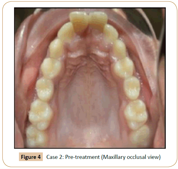
12 years 9 month old, growing female patient presented with a constricted maxillary arch, resulting in posterior crossbite and moderate crowding with proclined upper anteriors. Patient gave a history of thumbsucking habit till the age of 10 years which was discontinued spontaneously. Clinical examination showed some minor rotations with her upper and lower anterior teeth. Molar and canine relationship were Class I bilaterally (Figure 4).
Appliance design
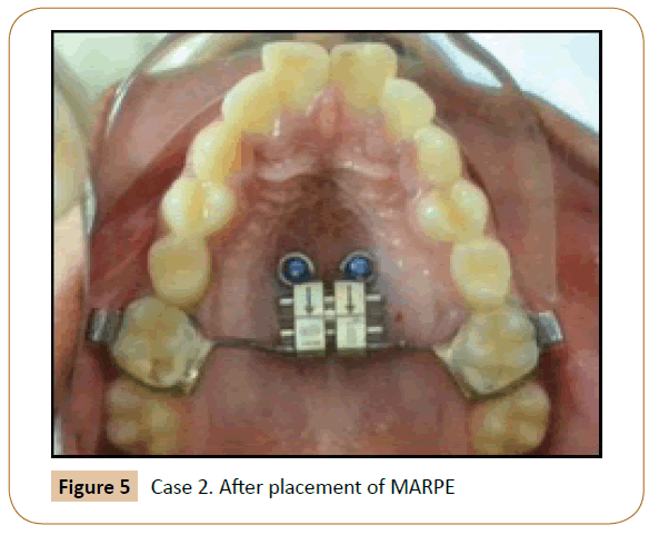
Anterior arms of the hyrax screw (Leone SPE, 9 mm) were bent in a circle to be used as implant supporting components. Two mini screws (1.8 x 10 mm, FavAnchorTM SAS, India) were placed in the paramedian region and the posterior arms of the appliance were anchored to the molars. Screw activation was performed as described (Figure 5).
Treatment progress
Treatment results: The maxillary intermolar and interpremolar width was as follows (Table 3 and Figure 6a-b).
| Pre-treatment | Post-expansion | Expansion | |
|---|---|---|---|
| Inter premolar | 39 mm | 41 mm | 2 mm |
| Inter molar | 50 mm | 55 mm | 5 mm |
Table 3: Case 2 Treatment results.
Case 3
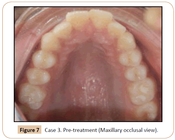
14 years 2 month old, growing female patient presented with forwardly placed maxillary anteriors and constricted maxillary arch. Clinical examination revealed mild proclination with upper and lower anteriors, increased overjet, erupting lower canines and class I molar relationship bilaterally (Figure 7).
Appliance design
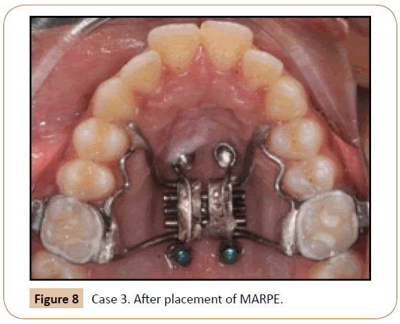
Four mini screws, two mini screws anterior (1.8 x 10 mm) and posterior (1.8 x 8 mm) each were placed in the paramedian region and the posterior arms of the appliance anchored to the molars and anterior arms extending on to the premolars (Figure 8).
Treatment progress
Treatment results: The maxillary intermolar and interpremolar width was increased by 6 mm (Pre-treatment: 54 mm; Post-treatment: 48 mm) and 3.5 mm (Pre-treatment: 38 mm; Posttreatment: 41.5 mm) respectively (Table 4 and Figure 9a-c).
| Pre-treatment | Post-expansion | Expansion | |
|---|---|---|---|
| Inter premolar | 38 mm | 41.5 mm | 3.5 mm |
| Inter molar | 48 mm | 54 mm | 6 mm |
Table 4: Case 3 Treatment results.
Discussion
In the present study, we evaluated skeletal and dentoalveolar changes after MARPE in young adults with transverse maxillary discrepancy. MARPE effectively achieved skeletal expansion by separation of the midpalatal suture.
Park et al. [20] reported a success rate of 84.2% in cases treated with a similar protocol. Skeletal expansion observed in the present study included the expansion of the zygomatic arch as well as nasal cavity. While zygomatic arch expanded to a lesser extent, expansion of nasal cavity was much more evident, and thus can result in improvement of nasal breathing owing to increased air flow. Thus, by effectively increasing the nasal cavity volume, treatment with a MARPE appliance can improve the constricted airway, thus aiding in long-term stability.
Garib G et al. [21], Gurgel JA et al. [22], also reported some amount of buccal tipping. Previous studies have reported greater changes in the degree of molar inclination than that of premolar inclination [15]. The higher density of the buccal cortical bone in the maxillary canine and premolar regions might have resulted in the greater buccal inclination of the first molar in comparison with that of the first premolar [20,23,24].
Using a MARPE appliance, some amount of buccal tipping is inevitable though much less as compared to RPE, as teeth are still used as anchor units alongside miniscrews. Tendency of buccal tipping is directly proportional to resistance exerted by midpalatal suture.
Conclusion
Expansion achieved in the cases treated by MARPE are majorly skeletal expansion, as the appliance is a tooth-and-tissue borne appliance. It can be used in young adults from late teens to mid-twenties and exhibits a high success in this particular age group [22,25,26].
Skeletal maxillary expansion was effectively accomplished using various designs of MARPE in all three of these patients. Clinical observations suggest that MARPE prevents many of the adverse effects of RPE and should be considered as a preferred and effective alternative for the same.
References
- Lee KJ, Park YC, Park JY, Hwang WS (2010) Mini screw assisted nonsurgical palatal expansion before orthognathic surgery for a patient with severe mandibular prognathism. Am J Orthod Dentofacial Orthop 137: 830-839.
- MacGinnis M, Chu H, Youssef G, Wu KW, Machado AW, et al. (2014) The effects of micro-implant assisted rapid palatal expansion (MARPE) on the nasomaxillary Complex: a finite element method (FEM) analysis. Prog Orthod 15: 52.
- Kim KB, Helmkamp ME (2012) Miniscrew implant-supported rapid maxillary expansion. J Clin Orthod 4: 608-612.
- Carlson C, Sung J, McComb RW, Machado AW, Moon W (2016) Microimplant-assisted rapid palatal expansion appliance to orthopedically correct transverse maxillary deficiency in an adult. Am J Orthod Dentofac Orthop 149: 716-728.
- Shapiro PA, Kokich VG (1988) Uses of implants in orthodontics. Dent Clin North Am 32: 539-550.
- Smalley WM, Shapiro PA, Hohl TH, Kokich VG, Branemark PI (1988) Osseointegrated titanium implants for maxillofacial protraction in monkeys. Am J Orthod Dentofacial Orthop 94: 285-295.
- Erverdi N, Okar I, Kucukkeles N, Arbak S (1994) A comparison of two different rapid palatal expansion techniques from the point of root resorption. Am J Orthod Dentofacial Orthop 106: 47-51.
- Parr JA, Garetto LP, Wohlford ME, Arbuckle GR, Roberts WE (1997) Sutural expansion using rigidly integrated endosseous implants: an experimental study in rabbits. Angle Orthod 67: 283-290.
- Harzer W, Reusser L, Hansen L, Richter R, Nagel T, et al. (2010) Minimally invasive rapid palatal expansion with an implant- supported hyrax screw. Biomed Tech (Berl) 55: 39-45.
- Marquezan M, Nojima LI, Freitas AOA, Baratieri C, Alves Jr M, et al. (2012) Tomographic mapping of the hard palate and overlying mucosa. Braz Oral Res 26: 36-42.
- Lombardo L, Gracco A, Zampini F, Stefanoni F, Mollica F (2010) Optimal palatal configuration for mini screw applications. Angle Orthod 80: 145-152.
- Kyung SH, Hong SG, Park YC (2003) Distalization of maxillary molars with a midpalatal mini screw. J Clin Orthod 37: 22-26.
- Kang S, Lee SJ, Ahn SJ, Heo MS, Kim TW (2007) Bone thickness of the palate for orthodontic mini-implant anchorage in adults. Am J Orthod Dentofac Orthop 131: 74-81.
- King KS, Lam EW, Faulkner MG, Heo G, Major PW (2007) Vertical bone volume in the paramedian palate of adolescents: a computed tomography study. Am J Orthod Dentofac Orthop 132: 783-788
- Moon SH, Park SH, Lim WH, Chun YS (2010) Palatal bone density in adult subjects: implications for mini-implant placement. Angle Orthod 80: 137-144.
- Kim HJ, Yun HS, Park HD, Kim DH, Park YC (2006) Soft-tissue and cortical-bone thickness at orthodontic implant sites. Am J Orthod Dentofac Orthop 130: 177-182.
- Motoyoshi M, Yoshida T, Ono A, Shimizu N (2007) Effect of cortical bone thickness and implant placement torque on stability of orthodontic mini-implants. Int J Oral Maxillofac Implants 22:779-784.
- Yılmaz A, Özçırpıcı AA, Erken S, Özsoy OP (2015) Comparison of short-term effects of mini- implant-supported maxillary expansion appliance with two conventional expansion protocols. Eur J Orthod 37: 556-64.
- Park JJ, Park YC, Lee KJ, Cha JY, Tahk JH, et al. (2017) Skeletal and dentoalveolar changes after mini screw- assisted rapid palatal expansion in young adults: A cone-beam computed tomography study. Korean J Orthod 47: 77-86.
- Garib DG, Henriques JF, Janson G, de Freitas MR, Fernandes AY (2006) Periodontal effects of rapid maxillary expansion with tooth-tissue-borne and tooth-borne expanders: a computed tomography evaluation. Am J Orthod Dentofacial Orthop 129: 749-758.
- Gurgel JA, Tiago CM, Normando D (2014) Transverse changes after surgically assisted rapid palatal expansion. Int J Oral Maxillofac Surg 43: 316-322.
- Christie KF, Boucher N, Chung CH (2010) Effects of bonded rapid palatal expansion on the transverse dimensions of the maxilla: a cone-beam computed tomography study. Am J Orthod Dentofacial Orthop 137: 79-85.
- Rungcharassaeng K, Caruso JM, Kan JY, Kim J, Taylor G (2007) Factors affecting buccal bone changes of maxillary posterior teeth after rapid maxillary expansion. Am J Orthod Dentofacial Orthop 132: 421-428.
- Corbridge JK, Campbell PM, Taylor R, Ceen RF, Buschang PH (2011) Transverse dentoalveolar changes after slow maxillary expansion. Am J Orthod Dentofacial Orthop 140: 317-325.
- Haas AJ (1970) Palatal expansion: just the beginning of dentofacial orthopaedics. Am J Orthod 57: 219- 255.
- Wehrbein H, Yildizhan F (2001)The mid-palatal suture in young adults. A radiological-histological investigation. Eur J Orthod 23: 105-114.
Open Access Journals
- Aquaculture & Veterinary Science
- Chemistry & Chemical Sciences
- Clinical Sciences
- Engineering
- General Science
- Genetics & Molecular Biology
- Health Care & Nursing
- Immunology & Microbiology
- Materials Science
- Mathematics & Physics
- Medical Sciences
- Neurology & Psychiatry
- Oncology & Cancer Science
- Pharmaceutical Sciences
