Interdisciplinary Approach in the Treatment of Peg Lateral Incisors
Rohit Kulshrestha
Saraswati Dental College and Hospital, Lucknow, India
- *Corresponding Author:
- Rohit Kulshrestha
BDS, MDS, Post Graduate Student
Saraswati Dental College and Hospital, Lucknow Orthodontics, India
Tel: +919870499761
E-mail: kulrohit@gmail.com
Received date: December 21, 2015; Accepted date: January 11, 2016; Published date: January 19, 2016
Citation: Kulshrestha R. Interdisciplinary Approach in the Treatment of Peg Lateral Incisors. J Orthod Endod. 2016, 2:1.
Abstract
Aberrations in tooth morphology resulting from late disturbances during the differentiation process most commonly result in size variations. Peg-shaped or mesio-distally deficient maxillary lateral incisors demonstrate variation in the expression of the trait, although the genes causing this are not known. Individuals with malformed lateral incisors often display a diastema in the midline region caused by the distal movement of the central incisor. These patients may exhibit otherwise normal dentitions unless other congenital etiologic factors or habits are present. The purpose of this article is to show an orthodontic and restorative approach for the treatment of peg-shaped lateral incisors.
Keywords
Peg lateral; Spacing; Composite veneering
Introduction
A peg lateral is defined as ‘‘an undersized, tapered, maxillary lateral incisor’’ [1] that may be associated with other dental anomalies, such as canine transposition and over retained deciduous teeth. Individuals with malformed lateral incisors often display a diastema in the midline region caused by the distal movement of the central incisor [2]. The incidence of peg-shaped incisors was found to be 0.8% in 739 Swedish children [3]. Arte et al [4] showed that the prevalence of hypodontia and/or peg-shaped teeth was over 40% in first- and second-degree relatives and 18% in first cousins in Finnish population. Four of nine noted obligate carriers of hypodontia gene had dental anomalies, including small upper lateral incisors, ectopic canines, taurodontism, and rotated premolars. They concluded hypodontia and/or peg shaped tooth was a genetic condition with autosomal-dominant transmission and that it was associated with several other dental abnormalities.
A rare disorder, oligodontia has always been considerably researched, especially concerning its prevalence and genetic background. Non-syndromic oligodontia has been associated with the presence of small and misshapen natural teeth, orofacial clefting and reduced saliva secretion [5]. Fraga et al [6] conducted a study to determine the relationship between toothsize reduction, agenesis and the occurrence of palatal displaced canines (PDCs). Emphasis was given to the association between anomalous and/or absent maxillary lateral incisors and PDCs. They found that association between tooth agenesis and PDCs was not observed. There also was no significant association between adjacent anomalous and/or absent maxillary lateral incisors and PDCs.
In the restoration of peg lateral incisors, there are many factors to be considered that depend on the patient’s expectations and the expertise of the clinician. The type of treatment should be selected based on functional and aesthetic requirements, need for extractions, the position of the canines and the potential for coordinating restorative and orthodontic treatment [7]. Treatment options vary but include the following [8]: (1) Extraction of the peg-shaped tooth and orthodontic movement of the canine into the space of the lateral incisor. The canines can then be re-contoured to resemble lateral incisors. (2) Extraction and replacement with a single-tooth implantsupported restoration or a fixed partial denture (FPD). (3) Direct or indirect restoration of the peg lateral incisors to develop normal tooth morphology after orthodontic alignment. Borzabadi-FarahanI [9] used implant-supported restorations in patients with hypodontia. It was challenging and required a multistage treatment that began with creating equal space between teeth followed by implant placement. Restorative treatment options include procedures such as porcelain laminate veneers, metal-ceramic restorations, and all-ceramic crowns, as well as minimally invasive procedures such as direct resin composite bonding veneers. It is essential that prior to any orthodontic treatment, full discussions are held with the restorative dentist, the patient, the patient’s parents and the orthodontist. This way all treatment options are fully discussed and outlined. The risks and benefits of all treatment can be discussed and outlined and any future long term restorative needs can be outlined. Timing and sequencing of appointments is essential towards the end of orthodontic treatment so that restorative treatment can be well coordinated at the correct appointment sequence [10]. This clinical report describes an interdisciplinary approach (Orthodontic and Restorative) for treatment of peg-shaped lateral incisors with a midline diastema.
Case Report
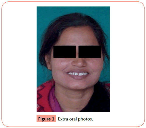
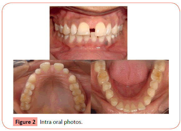
A female patient aged 26 came to the Department of Orthodontics with a chief complain of forwardly placed upper and lower front teeth with spacing. Medical history was non- relevant. A 4 mm midline diastema was seen on the frontal smiling photograph of the patient (Figure 1). On intra oral examination the patient had an Ellis Class 1 fracture with relation to upper left central incisor (Figure 2). Tongue thrusting habit was present. Peg lateral incisors were present in the maxillary arch. The mesio-distal dimensions of the peg laterals were reduced while the occluso-gingival height was normal. Spacing was seen in both arches, 12 mm in the maxillary arch and 3 mm in the mandibular arch. Mandibular midline was deviated to the right by 1mm. Class I molar and canine relation was seen. Cephalometric analysis readings are shown in Table 1.
| Sr. No | Parameter | Pre-treatment Value |
|---|---|---|
| 1 | Mandibular Plane Angle | 15 degrees |
| 2 | Incisor Mandibular Plane Angle | 126 degrees |
| 3 | Inter Incisal Angle | 102 degrees |
| 4 | Upper 1 to NA (angle) | 33 degrees |
| 5 | Upper 1 to NA (mm) | 10 mm |
| 6 | Lower 1 to NB (angle) | 43 degrees |
| 7 | Lower 1 to NB (mm) | 7 mm |
| 8 | Upper 1 to Point A | 8 mm |
| 9 | Lower 1 to A-Pog | 4 mm |
Table 1: Pre-treatment cephalometric values.
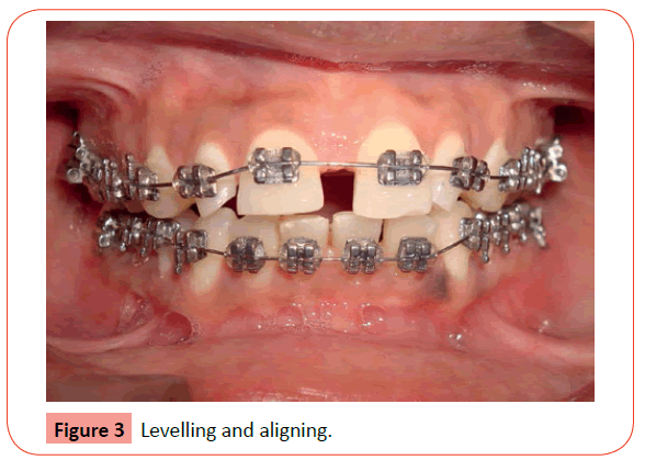
After mentioning all the treatment options to the patient she agreed upon the combined orthodontic and restorative treatment plan. The treatment plan was to orthodontically close the spaces in both the arches with sufficient space for veneer placement. Sufficient space (2 mm) was left between the maxillary incisors for the placement of composite veneers. The veneers would be placed from canine to canine so as to improve aesthetics and retention. The case was bonded with MBT Appliance 0.022 Slot and levelling and alignment was started with 0.016 NiTi wire (Figure 3).
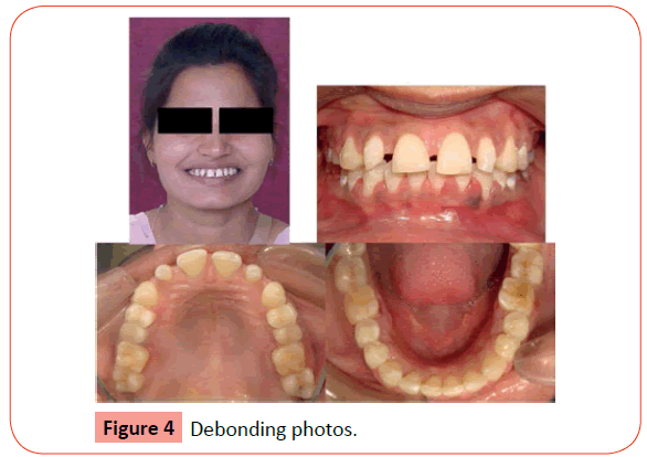
Levelling and alignment was completed in 6 months time. Once levelling was completed space closure was started. Lower arch space closure was completed in 4 months. 2 mm space was left between the maxillary incisors for veneering. Slight vertical discrepancy was seen with the incisal edges of the maxillary central incisors. This was due to the Ellis Class 1 fracture in relation with maxillary left central incisor. Debonding was done and the patient was referred to the Conservative Department for veneering preparation on the same day (Figure 4). Cephalometric values after debonding are shown in Table 2.
| Sr. No | Parameter | Post Treatment Value |
|---|---|---|
| 1 | Mandibular Plane Angle | 16 degrees |
| 2 | Incisor Mandibular Plane Angle | 120 degrees |
| 3 | Inter Incisal Angle | 112 degrees |
| 4 | Upper 1 to NA (angle) | 35 degrees |
| 5 | Upper 1 to NA (mm) | 8 mm |
| 6 | Lower 1 to NB (angle) | 32 degrees |
| 7 | Lower 1 to NB (mm) | 5 mm |
| 8 | Upper 1 to Point A | 6 mm |
| 9 | Lower 1 to A-Pog | 3 mm |
Table 2: Post-treatment cephalometric values.
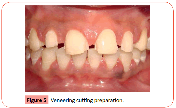
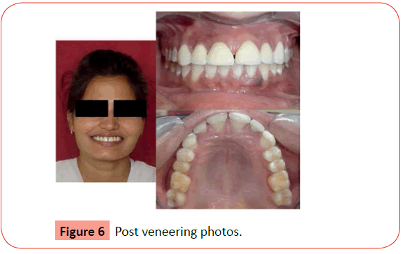
The incisor mandibular plane angle was reduced due to change in the mandibular incisor proclination. The maxillary and mandibular incisors were retracted by 2 mm each. The veneering cutting was done in a conservative manner (Figure 5). The veneers were luted on to the anteriors within 4 days (Figure 6). The midline in this case remained unchanged due to the fact that if the midline was corrected there would be unequal spaces between the incisors which in turn would affect the size and shape of the veneers also a midline discrepancy of 2-3 mm is acceptable.
Discussion
The aesthetic defect in patients with peg lateral incisors consists of both the malformed teeth and the presence of diastema between teeth. The treatment includes 2 primary objectives: to restore or replace the hypo plastic dental crowns or to close the diastema [11]. If the patient does not smoke or drink dark-coloured liquids that can alter the colour of the teeth, aesthetic bonding with resin composite may be the most conservative approach for several reasons: sound tooth structure will not be removed, the procedure may not require administration of local anaesthetic, the procedure may be accomplished in 1 appointment, and the treatment is relatively inexpensive [12].
Walls et al [13] used resin composite laminate veneers for masking discoloration or hypoplasia of the anterior teeth of 68 patients. The technique produced an acceptable improvement in the aesthetics and function of patients over a 2-year period.
The present case could have been treated directly by veneering. If direct veneering would have been done then only the spacing’s would have been closed, the proclination and forwardly placed incisors would not have been corrected. The chief complain of the patient was forwardly placed teeth followed by spacing; hence we treated the spacing problem later. Results of this clinical study showed that the gingival status of patients’ teeth improved significantly between the initial assessment visit and placement of the veneers. However, the veneer restorations showed a deleterious effect on the gingival health of the patients who were unable to maintain good oral hygiene.
Counihan [14] recommends that there are two basic approaches to treat peg lateral incisors. First, the lateral incisor can be extracted and the resultant space closed. However, this will often give a narrow anaesthetic smile. The second, preferred, option is often to open the space mesial and distal to the peg-lateral and create a proper space for a normal-sized lateral incisor. The restorative dentist has to build up the peg-lateral to simulate a normal-sized lateral incisor. It is best to discuss the positioning of the peg lateral within this space. Should it be close to the central incisor, in the middle of the space or to the distal of the space? This will depend on the actual size of the peg lateral and the amount of available space that can be created for the lateral incisor tooth.
Conclusion
The common occurrence of peg-shaped laterals is such that practitioners should be prepared for the careful interdisciplinary treatment planning to obtain excellent results. Through these interdisciplinary involvements with specialist colleagues a staged approach can be undertaken with orthodontic treatment being the first part of the treatment plan. This is followed by composite resin restorations to treat peg lateral teeth prior to the final retention phase of the orthodontic treatment. Appointments should be carefully co-ordinated so that this type of treatment can be efficiently and successfully carried out. The case presented here is just one example of how to treat peg lateral incisors with an interdisciplinary approach.
References
- Glossary of Prosthodontic Terms (1999) J Prosthet Dent81:90.
- Geiger A, Hirschfeld L (1974) Minor tooth movement in general practice. 3rd ed. St. Louis: Mosby 1-129, 349-434.
- Backman B, Wahlin YB (2001) Variations in number and morphology of permanent teeth in 7-year-old Swedish children. Int J Paediatr Dent 11:11-17.
- Arte S,Nieminen P,Apajalahti S,Haavikko K,Thesleff I,Pirinen S(2001)Characteristics ofincisor-premolar hypodontia in families. J Dent Res80:1445-1450.
- Kotsiomiti E,Kassa D,Kapari D (2007)Oligodontia and associated characteristics: assessment in view of prosthodontic rehabilitation. Eur J ProsthodontRestor Dent15:55-60.
- Fraga MR,Vitral RW,Mazzieiro ET (2012) Tooth size reduction and agenesis associated with palatal displaced canines. Pediatr Dent 34:216-219.
- Bello A, Jarvis RH (1997) A review of aesthetic alternatives for the restoration of anteriorteeth. J Prosthet Dent78:437-440.
- Contemporary Orthodontics - Proffit 4th ed. (2009) St Louis Mosby.
- Borzabadi-Farahani A (2012) Orthodontic considerations in restorative management of hypodontia patients with endosseous implants. Oral Implantol 38:779-791
- 10.Operative Dentistry - A Practical Guide to Recent Innovations - Hugh Devlin Springer.
- Textbook of Oral Pathology - Shafer 5th ed (2006) Elsevier.
- Studervants (2010) The Art and Science of Operative Dentistry - Studervant 5th ed Elsevier.
- Walls AW, Murray JJ, McCabe JF. (1988) Composite laminate veneers: A clinical study. J Oral Rehabil15:439-454.
- Counihan D (2000) The orthodontic restorative management of the peg lateral. Dental Update 27: 250-256.
Open Access Journals
- Aquaculture & Veterinary Science
- Chemistry & Chemical Sciences
- Clinical Sciences
- Engineering
- General Science
- Genetics & Molecular Biology
- Health Care & Nursing
- Immunology & Microbiology
- Materials Science
- Mathematics & Physics
- Medical Sciences
- Neurology & Psychiatry
- Oncology & Cancer Science
- Pharmaceutical Sciences
