Mini-invasive Orthodontic -surgical Treatment Of A Granuloma Localized Inside The Mandibular Canal.
G. Giordano, C. Luzi and C. M. A. Custode
G. Giordano1*, C. Luzi2 , C. M.A. Custode3
2A.M.C.O. (Rome) Department of Oral Surgery
3Prof. a.c. Ferrara University - Studio Luzi (Rome)
- *Corresponding Author:
- G. Giordano
A.M.C.O. (Rome) Department of Oral Surgery
Rome, Italy
Tel: +393473623431
E-mail: gianfrancogiordano@hotmail.it
Abstract
Proximity of the third mandibular molar to the mandibular canal generates risks of lesion to the mandibular nerve in case of surgical extraction procedures. The possibility of increasing the distance between the tooth and the nerve by means of orthodontic extrusion can be a valid strategic procedure to reduce the surgical risks. The authors applied a security protocol in a case of granuloma positioned in the mandibular canal in order to extrude and then extract tooth 4.8 with the aid of 2 TADs (Temporary Anchorage Devices) and a lever arm made of Titanium-Molybdenum-Alloy (beta-titanium).
Introduction
Various skeletal anchorage devices were introduced in the late 20th century for orthodontic purposes, including prosthodontic implants, zygoma ligatures, palatal onplants and implants, retro molar implants, miniplates, and surgical screws. The latter, which became known as temporary anchorage devices (TADs), have become increasingly popular because they are small and easy to insert and remove, they can be loaded immediately after insertion, and they can provide absolute anchorage for many types of orthodontic treatment, with no need for special patient compliance [1-5].
Absolute anchorage control is today achievable for many daily different clinical challenges. The possibility of generating force systems that do not rely on neighboring teeth has made orthodontic treatment a valid solution for many adult patients presenting extensive prosthodontics, periodontal problems and complicated severe malocclusions. For these patients skeletal anchorage, in conjunction with appropriate biomechanics, has widened the spectrum of the clinical possible treatments. The case report describes a technique the authors have set-up for the extrusion of a lower third molar, presenting a peri-apical granuloma and in close proximity with the mandibular canal, in order to perform consecutively a safe extraction procedure.
Materials and methods
A 33 year-old female patient presented noticeable swelling of the cheek and difficulty opening her mouth. The patient reported a toothache located at the level of the tooth 4.8. The lower third of the patient’s face appeared reduced.
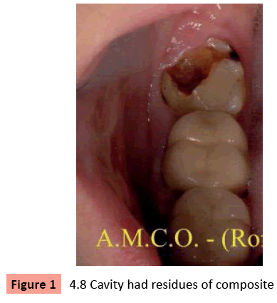
On examination, the patient presented many incongruous prosthetic replacements as well as infiltrated reconstructions. Much of the clinical crown of the element appeared to be destroyed (Figure 1).
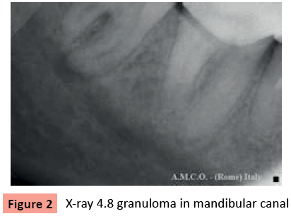
The cavity had residues of composite material from a previous reconstruction as well as organic, malodorous material. An intraoral X-ray (Figure 2) of the element revealed what seemed to be an apical granuloma in the mandibular canal. This conclusion was confirmed some performing a 3-D cone days later after beam (Figure 3) image reconstruction.
Given the clinical evidence and the suspicious granuloma (the cause leading to the formation of granuloma is formed by bacterial toxins and necrosis of the dental pulp ["nerve"] that remain in the root canals untouched by the instruments or disinfectants) jutting into the mandibular canal, the risk of compromising the perineurium if the patient was submitted to a normal tooth extraction arose. The alternative was to perform a piezo-surgery; an option which was discarded immediately since the patient was uncomfortable with the destructive effects that this procedure may have caused to the mandible. In order to avoid the risk of permanent paresthesia or an incremental hemiparesis, it was decided to use two TADs along with a lever to determine the orthodontic disinclusion or the gradual detachment of the eighth position.
Antibiotic therapy was prescribed as required by international protocol; (amoxicillin 875 mg, and potassium clavulanate equivalent to clavulanic acid 125 mg [medication brand name, Clavulin] 1 tablet every 12 hours for 7 days, naproxen sodium 550 mg [medication brand name, Synflex 550mg ] 1 capsule twice daily for the first 3 days and then with a meal once a day for another 4 days; Omeprazole 20 mg [medication brand name , Antra] 1 capsule in the morning; probiotic [ferment lactic eptaceppo with vitamin B-complex: Code AIC: 906419989source:https://www. fogliettoillustrativo.net/906419989/floragen-fermenti-lattici- 30pz#.VE1Qtcm2Afg molecular name, Floragen] 1-2 capsules three times a day. In addition, the cavity was cleaned and treated with a eugenol based medication after administrating nerve block anesthesia.
As previously stated, a three-dimensional image reconstruction confirmed the presence of an apical granuloma projecting towards the mandibular canal. The reconstruction was done using the NobelClinician™ cone beam made with newton 5 and axial images. The authors decided to continue the antibiotics previously prescribed and proceed with a pulp devitalization. During the procedure, the authors made sure to stay about 3 mm deep of the apexes due to the fact that the measurements made with both orthopanoramic and with both the cone beam indicated that the apex of the distal root seemed to jut into the mandibular canal about 1 mm. The depth control was done both radiologically and by using an ERCLMD [Electronic Root Canal Length Measurement] device.
Once the root canal was completed (11/12/2013), the next step was to position two TADs (Aarhus mini-implant 6mm thread length and 1.5mm diameter) (11/25/13) at the level of the adjacent gingivae (second protocol Aarhus); the most mesial end at the level of 4.6 and the most distal end of the fork positioned between 4.7 and 4.8.
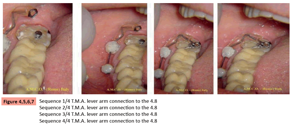
The authors positioned (Figures 4-7) the beta-titanium lever arm and deferred the connection to the 4.8 element (11/27/2013) to avoid contamination of the site. The estimated time for the lever to serve its purpose was 6 months.
Subsequently, the authors proceeded with the reconstruction of the crown. An orthodontic button was inserted into the composite material. The lever arm was attached to the TAD with a metal ligature coated with composite to prevent bedsores. Also the angle of the lever was coated with a protective silicone material.
The lever was activated on a monthly basis, and each time the element was checked radiologically, as per protocol.
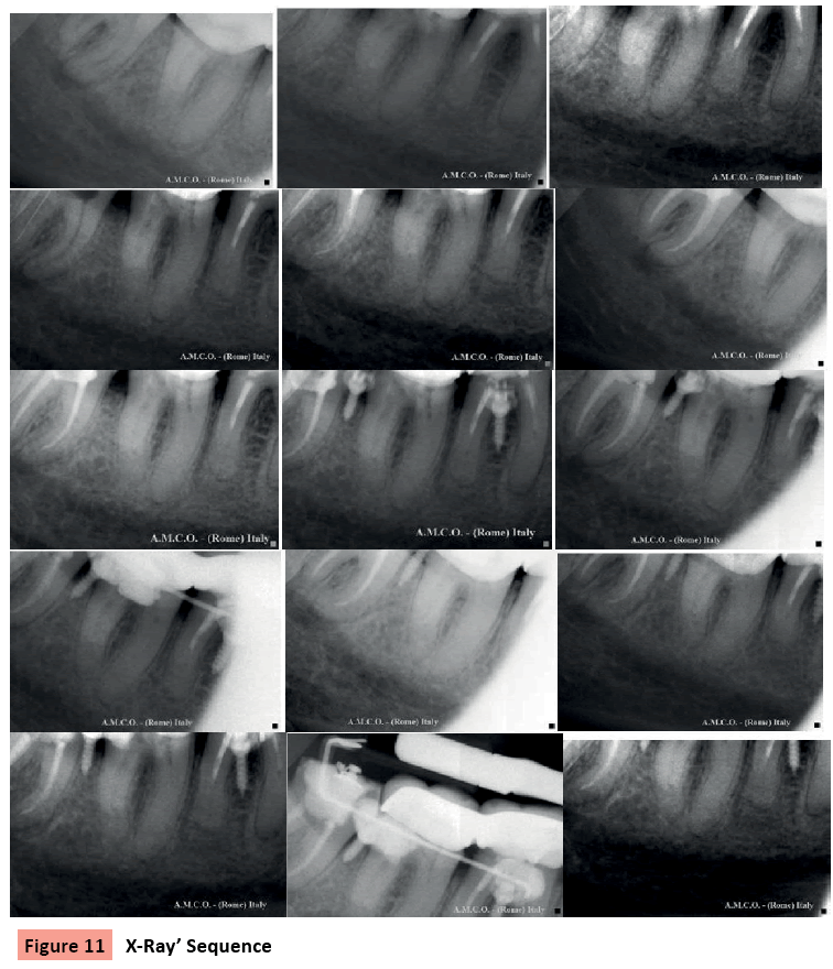
After two months the authors replaced the silicone protection(Figure 8); radiological controls showed [Rx 'Sequence] a gradual lifting of the element on a monthly basis. Each month the crown was milled to gradually reduce it.
After five months, radiology showed an evident removal of both the quotes and related granuloma from the mandibular canal. This removal occurred one month before the estimated date. The authors then decided to remove the lever arm and the two TADs as well as to extract the 4.8 element which was safe to remove at this stage.
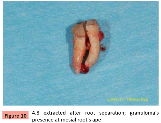
The surgery began on 04/22/2014 [Surgery's Day Pics] with the release of the lever arm from both the orthodontic button and the TADs, followed by the removal of the TADs. The element was sectioned using a diamond bur and then extracted and the mesial root brought out the granuloma.
The element was entirely rebuilt on the operating table and the vacuum was controlled through radiography.
The patient awoke from her anesthesia nerve block without any symptoms of paresthesia. A follow up was done three months after surgery and the patient appeared perfectly healed.
Conclusions
One of the major challenges of oral surgery is the surgical extraction of a lower third molar with the roots in proximity to the mandibular canal. Furthermore, the presence of an apical granuloma is a complication that every surgeon would like to avoid. The use of a lever arm with mini screw anchorage under a security protocol may facilitate the surgical removal of the tooth by moving away its apex and granuloma from the mandibular canal. This technique also reduces the discomfort of the patient who does not undergo the stress of invasive techniques for the extraction of the element or its side effects. This safety technique eliminates the possibility of a serious permanent injury that could complicate an already difficult surgery.
The authors declare that there is no conflict of interests regarding the publication of this article.
References
- Costa A, Raffaini M, and Melsen B. (1998): Miniscrews as orthodontic anchorage: A preliminary report, Int. J. Adult Orthod. Orthog. Surg. 13: 201-209.
- Kyung, H.M, Park H.S, Bae S.M, Sung J.H and Kim (2003) I.B.: Development of orthodontic micro-implants for intraoral anchorage J. Clin. Orthod. 37: 321-328.
- Carano, A, Velo S, Incorvati C and Poggio P(2004) Clinical applications of the Mini-Screw-Anchorage-System (M.A.S.) in the maxillary alveolar bone, Prog. Orthod. 5:212-235.
- Creekmore T D and Eklund M K (1983) The possibility of skeletal anchorage . Journal of Clinical Orthodontics 17: 266 – 269.
- Costa A , Raffaini M , Melsen B (1998) Miniscrews as orthodontic anchorage: a preliminary report . International Journal of Adult Orthodontics and Orthognathic Surgery 13: 201 – 209.
Open Access Journals
- Aquaculture & Veterinary Science
- Chemistry & Chemical Sciences
- Clinical Sciences
- Engineering
- General Science
- Genetics & Molecular Biology
- Health Care & Nursing
- Immunology & Microbiology
- Materials Science
- Mathematics & Physics
- Medical Sciences
- Neurology & Psychiatry
- Oncology & Cancer Science
- Pharmaceutical Sciences
