Minimal Management Of Traumatically Luxated Immature Maxillary Permanent Incisors
Peter M. Di Fiore
Peter M. Di Fiore*
Diplomate, American Board of Endodontics Professor, Department of Endodontics University of Texas, School of Dentistry, Houston, Texas
- *Corresponding Author:
- Dr. Peter M. Di Fiore
University of Texas, School of Dentistry, Department of Endodontics
7500 Cambridge St. Ste. 6400, Houston, TX 77054, United States
E-mail: petermdifiore@aol.com
Abstract
Background: Luxations account for half of all traumatic dental injuries and occur most frequently to maxillary incisors during childhood and adolescence. The dental pulp response to traumatic luxation injuries can be survival, calcification or necrosis, depending on the severity and type of injury and the developmental stage of the tooth. However, the dental pulps of severely luxated immature teeth have the ability to remain viable and continue to function.
Methods: An 8 year old girl sustained traumatic luxation injuries of different types to her maxillary immature permanent incisors that were repositioned and stabilized for 1 month and managed without endodontic intervention. Clinical and radiographic follow-up examinations were conducted at 6-month intervals for 2 ½ years.
Findings: Assessment at the end of the follow-up period revealed continued root growth and apical development, the absence of pulpal and periapical disease and retention of the maxillary incisors in a state of health and normal function.
Conclusions: Careful and continual assessment is necessary to determine the progress and prognosis of pulpal healing for traumatically luxated immature teeth.
Introduction
In dentistry tooth luxation is defined as the partial separation of a tooth from its socket [1]. The World Health Organization classification of traumatic dental injuries subdivides luxation injuries, into 5 types: concussions, subluxations and extrusive, intrusive and lateral luxations [2]. Traumatic injuries to permanent teeth, of which approximately 55% are luxations, predominantly occur during the 10 year period of 6 to 16 years of age and overwhelmingly affect the maxillary incisors [3-10].
The dental pulp response to tooth luxation can have essentially three outcomes: survival, calcification or necrosis, depending upon the severity and type of injury and the stage of tooth development [11-17]. Pulp survival is maintained to a high degree with mild to moderate luxation injuries (concusions and subluxations), while with severe luxation injuries (extrusive, intrusive and lateral luxations) pulp necrosis is a frequent occurrence [11-17]. However, the dental pulps of severely luxated immature teeth have a much higher rate of survival than those of mature teeth [11-13,17].
An investigation of the prognosis of 637 luxated permanent teeth found that pulp necrosis occurred in 3% of concussions, 6% of subluxations, 26% of extrusive luxations, 58% of lateral luxations and 85% of intrusive luxations. However the differences in the occurrence of pulp necrosis among mature versus immature teeth were respectively; 4% to 0% for concussions, 15% to 0% for subluxations, 64% to 9% for extrusive luxations, 77% to 9% for lateral luxations and 100% to 65% for intrusive luxations [12].
Another investigation of 226 luxated teeth that correlated the dimension of the apical foramen with the occurrence of pulp necrosis over a 10 year period, showed that pulp necrosis was significantly related to the size of the apical foramen, with a higher incidence of pulp necrosis occurring in teeth with a smaller apical foramen and a lower rate of pulp necrosis in teeth with a larger apical foramen [18].
Contemporary treatment guidelines for the management of luxated permanent teeth are based on the type and severity of the injury and the stage of tooth development at the time of injury, and generally recommend early pulp removal for severely luxated mature teeth where there is a higher incidence of pulp necrosis due to pulpal ischemia, and pulp retention for severely luxated immature teeth where there is a lower incidence of pulp necrosis due to the presence of a rich vascular blood supply to the pulp through a wide open apical foramen that maintains pulpal circulation [19,20].
This is a clinical case study of an 8 year old child who sustained different types of traumatic luxation injuries to her immature maxillary permanent incisors, that were managed without endodontic intervention and were continually assessed on a 6 month basis for 2 ½ years. The teeth continued to develop and mature and were retained in a state of health and normal function.
Case Report
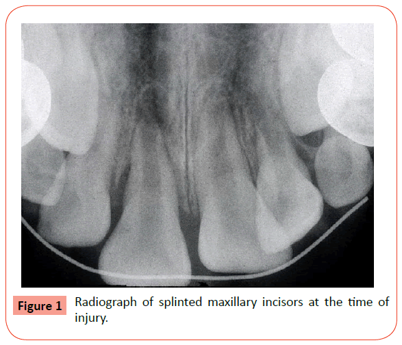
An 8 year old girl had presented with her mother to a dental clinic for after-hours emergency treatment of dental injuries sustained when she accidentally fell while riding a skateboard and her mouth hit the sidewalk pavement. It was reported, on examination by the on-call attending dentist, that the child had sustained abrasions and contusions of her upper and lower lips and that the right maxillary lateral incisor had been loosened, the right maxillary central incisor extruded, the left maxillary central incisor intruded and the left lateral incisor palatally luxated and severely loosened. The abrasions and contusions of the lips were treated. Repositioning of the teeth was attempted with limited success and the teeth were splinted with composite bonded ligature wire placed across the anterior arch from the right to the left deciduous canines and a radiograph was taken (Figure 1).
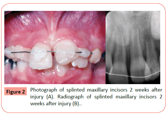
One week after injury, the patient presented for endodontic evaluation of her traumatized maxillary incisor teeth. Clinical examination revealed an apprehensive and anxious but healthy 8 year old girl with a non-contributory medical history, no known allergies and generalized dental plaque accumulation. Healing of the lip abrasions and contusions was observed. The maxillary incisors were mal-aligned, the ligature wire was unattached at the lateral incisors, and the left lateral incisor was mobile (Figure 2A). The ligature wire was re-bonded to the lateral incisors with composite resin and a radiograph was taken which revealed the splinted immature incisors with traumatically induced periapical rarefactions (Figure 2B). The patient received oral hygiene instructions and was reappointed for follow-up examination. A diagnostic summary of the different types of traumatic luxation injuries sustained were: subluxation of the maxillary right lateral incisor, extrusive luxation of the maxillary right central incisor, intrusive luxation of the maxillary left central incisor and lateral luxation with mobility of the maxillary left lateral incisor.
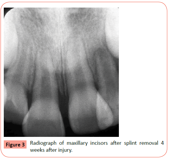
One month after injury, the patient was asymptomatic, the lip abrasions and contusions were completely healed, the splint was removed and the teeth were stable. The gingival attachments were intact and the alveolar mucosa was normal without pain or tenderness to palpation. Radiographic examination showed immature incisors with resolution of the traumatically induced transient periapical rarefactions (Figure 3). The patient received a dental cleaning and tooth brush instructions. Appointments for subsequent follow-up examinations and dental cleanings were scheduled on a 6 month post-injury basis.
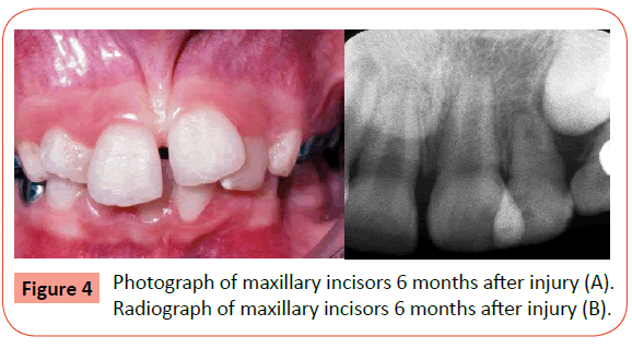
Six months after injury the patient was asymptomatic. Clinical examination revealed stable maxillary incisors with normal mobility, intact gingival attachments, no discoloration and no pain on percussion or tenderness to palpation (Figure 4 A). Pulp sensibility tests were positive for all of the maxillary incisors except for the left lateral incisor. Radiographic examination of the maxillary incisors revealed no periapical or periradicular radioliucencies, continued root growth and apical development for all of the maxillary incisors except for the left lateral incisor which showed delayed root growth and pulpal calcification (Figure 4 B).
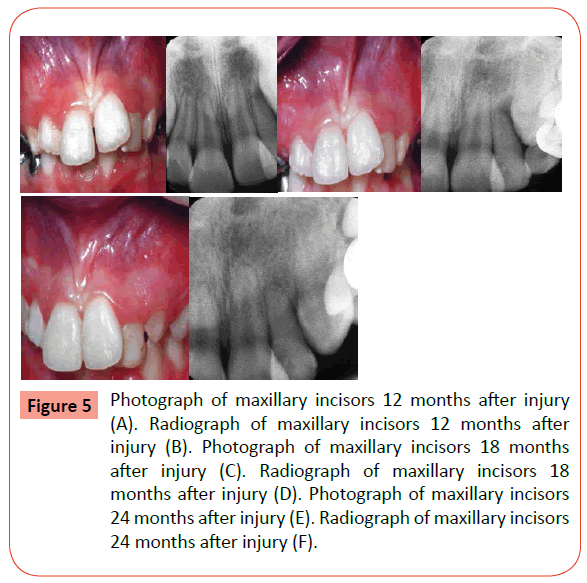
Clinical and radiographic examinations, 12 months after injury (Figure 5 A and B), 18 months after injury (Figure 5 C and D) and 24 months after injury (Figure 5 E and F), revealed that the teeth were stable with normal mobility and intact gingival attachments. There was no pain on percussion or tenderness to palpation, and except for the left lateral incisor which showed slight discoloration and was non-responsive to pulp sensibility tests, all of the others incisors continued to have positive responses to pulp sensibility tests and no discoloration. The midline diastema had closed and the alignment of the central incisors had improved. The left permanent canine continued to descend and erupt into position as the left deciduous canine exfoliated. Root growth and apical development had progressed for the right lateral, right central and left central incisors while the left lateral incisor showed arrested root development and pulp canal obliteration. The periapical and periradicular bone of all the maxillary incisors appeared within normal limits at all 3 follow-up examinations.
Figure 5: Photograph of maxillary incisors 12 months after injury (A). Radiograph of maxillary incisors 12 months after injury (B). Photograph of maxillary incisors 18 months after injury (C). Radiograph of maxillary incisors 18 months after injury (D). Photograph of maxillary incisors 24 months after injury (E). Radiograph of maxillary incisors 24 months after injury (F).
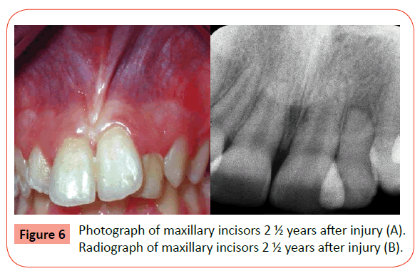
At the final recall examination 2 ½ years after the injury, with the patient then at 10 ½ years of age, the maxillary incisors were asymptomatic and functioning normally and all except for the left lateral incisor, which had undergone complete pulp calcification, responded normally to pulp sensibility tests. The surrounding oral tissues were normal and the left permanent canine had erupted into its normal position in the maxillary anterior arch (Figure 6 A). Radiographic examination revealed normal appearing maxillary anterior alveolar bone with no periapical and periradicular radiolucencies. The right lateral incisor and the right and left central incisors showed continued root growth and apical development while the left lateral incisor showed arrested root growth with blunted apical development and total pulp canal obliteration (Figure 6B).
Discussion
This was a clinical case of dental trauma in an 8 year old girl, who sustained luxation injuries of different types to each of her immature maxillary permanent incisors. The teeth were initially repositioned and stabilized with a flexible composite bonded wire splint four 4 weeks and then minimally managed, without endodontic intervention, and continuously monitored at intervals of 6 month after injury for a follow-up period of 2 ½ years. Flexible wire-composite splints [21] of the type applied in this case can be left in place for an extended time without any ill effects for periodontal ligament healing, since they allow for normal functional tooth movement [22-25]. In vitro and in vivo investigations, that measured the lateral tooth movement of teeth with this type of splint, have demonstrated that its immobilization effect did not exceed normal tooth firmness and that it provided a degree of tooth mobility similar to non-splinted teeth [22-25]. Therefore, the longer splinting time of 4 weeks as opposed to the standard of 2 weeks for these luxated immature teeth was appropriate and reasonable.
At 4 weeks after injury the findings of non-responsive pulp sensibility test results were not considered to be conclusive for developing traumatized teeth, since there can be a temporary loss of sensory nerve function which can later return to normal [26-29].
Additionally, pulp vitality test results with immature teeth can be misleading [30,31] because full development of the Plexus of Raschkow and full pulpal innervation with A-delta nerve fibers takes place after root formation has been completed [32].
Radiographic examination of the traumatized immature maxillary incisors at 4 weeks after injury revealed the presence of traumatically induced transient periapical rarefactions which later completely resolved at 8 weeks. This finding was consistent with observations among luxated permanent teeth of transient apical breakdown that appeared spontaneously as periapical radiolucencies then later resolved to normal periapical radiographic appearances [33].
Oral hygiene was emphasized and tooth brush instruction was given since the patient had heavy dental plaque accumulation. The teeth were then carefully assessed at each of the 6 month follow visits for a period of 2 ½ years and the child also received regular dental cleanings.
At the 6 month follow-up assessment pulp sensibility tests were normal for all of the maxillary incisors with the exception of the left lateral incisor which radiographically showed pulpal canal obliteration. Traumatically induced dental pulp calcification is not an unusual finding and can be considered a type of healing response to pulpal injury [13-17].
The incidence of occurrence of pulp calcification among 637 permanent teeth that sustained luxation type injuries was found to be 15% (96/637) of which only 1 developed pulpal necrosis, suggesting that this type of calcification may be a physiological rather than a pathologic response [13]. Among 122 traumatized permanent anterior teeth with calcifying pulps that were followed over a long time-period of 10-23 years, periapical pathosis was observed in 13% of all teeth, 17% of teeth with narrow apical foramen and only 5% of teeth with wide apical foramen [34].
Since the clinical and radiographic examination findings at 6 months after injury for all of the maxillary incisors were normal with the exception of the negative pulp sensibility test for the left lateral incisor with pulpal calcification, the decision was made to carefully follow these teeth and allow the dental pulps to continue with dentin formation for further root growth and apical development.
Each of the subsequent follow-up examinations at 12, 18 and 24 months after the injury showed that the traumatized teeth continued to progress in their development without any adverse signs or symptoms of pulpal or periapical inflammatory disease.
At the final recall examination, 2 ½ years after injury, the right lateral incisor which had been subluxated and sustained the mildest injury, showed normal root development, while the right central incisor which had been extruded and left central incisor which had been intruded showed a slightly aberrant root development pattern and a mild degree of surface root resorption. The left lateral incisor which had been laterally luxated and severely mobilized showed arrested root growth and total pulp calcification. Several clinical studies that investigated the types of healing responses that can occur as sequelae to periodontal and pulpal injury of traumatically luxated teeth, found post-traumatic conditions similar to those found in this case [13-17].
This was the overall outcome in the case presented for these traumatized immature maxillary permanent incisors that had sustained different types of luxation injuries and were assessed for 2 ½ years using standard clinical and radiographic criteria to determine their pulpal, periapical and periodontal status [35]. All four incisors were eventually retained in a state of health and normal function, and although they were mal-aligned, orthodontic treatment later corrected their position in the maxillary anterior dental arch.
The severity and type of the luxation injuries are critical factors for pulp survival in traumatically luxated teeth [11-17]. In a 3 part investigation on the outcomes of 144 severely luxated permanent incisors in patients whose ages ranged from 6 to 18 years with a mean age of 10 years, and who were followed over a mean period of 3.8 years, the rates of occurrence for pulp necrosis, pulp survival and pulp calcification were 43%, 34% and 23% respectively [36-38]. However, the pulps of traumatically luxated immature teeth have been shown to have a much higher rate of pulp survival and a much lower rate of pulp necrosis than mature teeth with every type of luxation injury [11-13,17]. Clinical assessments of the prognosis for survival of the dental pulps of 637 luxated permanent teeth, found that 9% of the teeth with closed apices developed pulp necrosis after mild to moderate luxation injuries and 80% developed pulp necrosis after severe luxation injuries, while none of the teeth with open apices developed pulp necrosis after mild to moderate luxation injuries and the development of pulp necrosis significantly decreased to only 23% of the teeth that sustained severe luxation injuries [12,13].
In an investigation of the pulpal status of luxated teeth, it was found that on the initial examination of 26 teeth with discoloration and loss of sensibility as possible clinical indications of pulpal necrosis, all showed normal coloration and sensibility on subsequent examination and that among the extirpated pulps of 66 luxated teeth in which pulpal necrosis was deemed to have occurred, 12 histologically revealed the presence of viable cells and blood vessels, indicating a pulpal healing response [39].
A follow-up investigation of the pulpal and periodontal healing of 37 laterally luxated permanent teeth at 4 years post-trauma found that among teeth with closed apices 9 developed pulp necrosis and 1 developed external root resorption, but among teeth with open apices none developed either pulp necrosis or external root resorption, indicating a favorable prognosis for pulp survival and periodontal healing in laterally luxated immature teeth [40].
Longitudinal cohort studies of 469 concussed, 404 subluxated , 82 extruded and 179 laterally luxated teeth which were followed at regular intervals of weeks, months and years, determined that after 3 years the risk of periodontal ligament healing complications was generally low for mature teeth and very low for immature teeth [41,42].
Of all the types of luxation injuries, intrusive luxations are the most damaging to the apical tissues, the dental pulp and the periodontal ligament, and have the highest incidence of pulp necrosis and root resorption [11]. However the pulps of luxated teeth with incomplete root formation have a higher rate of survival than those with complete root formation and closed apices [43]. There is also, depending upon the degree of intrusion, the opportunity for the pulps intruded teeth to heal, especially if the intrusion is less than 3mm [36]. In two retrospective studies of patients, in similar age brackets of (6 to 16 and 6 to 17 years), with a combined sample of 111 intrusively luxated incisors, 52 teeth spontaneously re-erupted and continued to develop and it was concluded that allowing for re-eruption without any active treatment resulted in less complications for intruded immature incisors [44,45].
These clinical investigations [36-45] on the outcomes of various types of luxation injuries showed that for immature permanent teeth the opportunity for pulpal and periodontal healing, continued root development and apical maturation was favorable and that the prognosis for retention without endodontic intervention was good.
Conclusion
This clinical case report served to demonstrate that careful and continual assessment was necessary to follow the progress and determine the prognosis for a favorable healing outcome of traumatically luxated immature permanent maxillary incisors. The primary approach in the management of children and adolescents who sustain traumatic luxation injuries should be directed toward supporting the healing potential that is naturally inherent in their developing dentoalveolar tissues. Endodontic treatment interventions would be indicated only when there are clinical and radiographic signs of pulpal and periapical inflammatory pathosis.
Acknowledgement Disclosure Statement
The author has no conflicts of interest.
References
- Stedman’s Medical Dictionary, Lippincott, Williams & Wilkins, 27th edn. 2000.
- Application of the International Classification of Diseases to Dentistry and Stomatology. ICD-DA 3rd edn. World Health Organization, Geneva 1995.
- Andreasen JO (1970) Etiology and pathogenesis of traumatic dental injuries. A clinical study of 1,298 cases. Scand J Dent Res 78: 329-342.
- Oikarinen K, Kassila O (1987) Causes and types of traumatic tooth injuries treated in a public dental health clinic. Endod Dent Traumatol 3: 172-177.
- Oikarinen K (1987) Pathogenesis and mechanism of traumatic injuries to teeth. Endod Dent Traumatol 3: 220-223.
- Zerman N, Cavalleri G (1993) Traumatic injuries to permanent incisors. Endod Dent Traumatol 9: 61-64.
- Schatz JP, Joho JP (1994) A retrospective study of dento-alveolar injuries. Endod Dent Traumatol 10: 11-14.
- Luz JG, Di Mase F (1994) Incidence of dentoalveolar injuries in hospital emergency room patients. Endod Dent Traumatol 10: 188-190.
- Skaare AB, Jacobsen I (2003) Dental injuries in Norwegians aged 7-18 years. Dent Traumatol 19: 67-71.
- Crona-Larsson G, Norén JG (1989) Luxation injuries to permanent teeth -- a retrospective study of etiological factors. Endod Dent Traumatol 5: 176-179.
- Andreasen JO (1970) Luxation of permanent teeth due to trauma. A clinical and radiographic follow-up study of 189 injured teeth. Scand J Dent Res 78: 273-286.
- Andreasen FM, Pedersen BV (1985) Prognosis of luxated permanent teeth--the development of pulp necrosis. Endod Dent Traumatol 1: 207-220.
- Andreasen FM, Yu Z, Thombsen B, Andersen PK. (1987) Occurrence of pulp canal obliteration after luxation injuries in the permanent dentition. Endod Dent Traumatol 3: 103-105.
- Oikarinen K, Gundlach KK, Pfeifer G (1987) Late complications of luxation injuries to teeth. Endod Dent Traumatol 3: 296-303.
- Andreasen FM (1989) Pulpal healing after luxation injuries and root fracture in the permanent dentition. Endod Dent Traumatol 5: 111-131.
- Crona-Larsson G, Bjarnason S, Norén JG (1991) Effect of luxation injuries on permanent teeth. Endod Dent Traumatol 7: 199-206.
- Feiglin B (1996) Dental pulp response to traumatic injuries--a retrospective analysis with case reports. Endod Dent Traumatol 12: 1-8.
- Andreasen FM, Zhijie Y, Thomsen BL (1986) Relationship between pulp dimensions and development of pulp necrosis after luxation injuries in the permanent dentition. Endod Dent Traumatol 2: 90-98.
- Flores MT, Andersson L, Andreasen JO, Bakland LK, Malmgren B, et al. (2007) Guidelines for the management of traumatic dental injuries. I. Fractures and luxations of permanent teeth. Dent Traumatol 23: 66-71.
- Diangelis AJ, Andreasen JO, Ebeleseder KA, Kenny DJ, Trope M, et al. (2012) International Association of Dental Traumatology guidelines for the management of traumatic dental injuries: 1. Fractures and luxations of permanent teeth. Dent Traumatol 28: 2-12.
- Oikarinen K (1990) Tooth splinting: a review of the literature and consideration of the versatility of a wire-composite splint. Endod Dent Traumatol 6: 237-250.
- Oikarinen K (1987) Functional fixation for traumatically luxated teeth. Endod Dent Traumatol 3: 224-228.
- Oikarinen K (1988) Comparison of the flexibility of various splinting methods for tooth fixation. Int J Oral Maxillofac Surg 17: 125-127.
- Oikarinen K, Andreasen JO, Andreasen FM (1992) Rigidity of various fixation methods used as dental splints. Endod Dent Traumatol 8: 113-119.
- Ebeleseder KA, Glockner K, Pertl C, Städtler P (1995) Splints made of wire and composite: an investigation of lateral tooth mobility in vivo. Endod Dent Traumatol 11: 288-293.
- Skieller V. (1960) The prognosis for young teeth loosened after mechanical trauma. Acta Odont Scand. 18: 171-81.
- Teitler D, Tzadik D, Eidelman E, Odont, Chosack A (1972) A clinical evaluation of vitality tests in anterior teeth following fracture of enamel and dentin. Oral Surg Oral Med Oral Pathol 34: 649-652.
- Rock WP, Grundy MC (1981) The effect of luxation and subluxation upon the prognosis of traumatized incisor teeth. J Dent 9: 224-230.
- Bastos JV, Goulart EM, de Souza Côrtes MI (2014) Pulpal response to sensibility tests after traumatic dental injuries in permanent teeth. Dent Traumatol 30: 188-192.
- Fulling HJ, Andreasen JO (1976) Influence of maturation status and tooth type of permanent teeth upon electrometric and thermal pulp testing. Scand J Dent Res 84: 286-290.
- Klein H (1978) Pulp responses to an electric pulp stimulator in the developing permanent anterior dentition. ASDC J Dent Child 45: 199-201.
- Harris R, Griffin CJ (1968) Fine structure of nerve endings in the human dental pulp. Arch Oral Biol 13: 773-779.
- Andreasen FM (1986) Transient apical breakdown and its relation to color and sensibility changes after luxation injuries to teeth. Endod Dent Traumatol 2: 9-19.
- Jacobsen I, Kerekes K (1977) Long-term prognosis of traumatized permanent anterior teeth showing calcifying processes in the pulp cavity. Scand J Dent Res 85: 588-598.
- Jacobsen I (1980) Criteria for diagnosis of pulp necrosis in traumatized permanent incisors. Scand J Dent Res 88: 306-312.
- Humphrey JM1, Kenny DJ, Barrett EJ (2003) Clinical outcomes for permanent incisor luxations in a pediatric population. I. Intrusions. Dent Traumatol 19: 266-273.
- Lee R, Barrett EJ, Kenny DJ (2003) Clinical outcomes for permanent incisor luxations in a pediatric population. II. Extrusions. Dent Traumatol 19: 274-279.
- Nikoui M, Kenny DJ, Barrett EJ (2003) Clinical outcomes for permanent incisor luxations in a pediatric population. III. Lateral luxations. Dent Traumatol 19: 280-285.
- Andreasen FM (1988) Histological and bacteriological study of pulps extirpated after luxation injuries. Endod Dent Traumatol 4: 170-181.
- Ferrazzini Pozzi EC1, von Arx T (2008) Pulp and periodontal healing of laterally luxated permanent teeth: results after 4 years. Dent Traumatol 24: 658-662.
- Hermann NV, Lauridsen E, Ahrensburg SS, Gerds TA, Andreasen JO (2012) Periodontal healing complications following concussion and subluxation injuries in the permanent dentition: a longitudinal cohort study. Dent Traumatol 28: 386-393.
- Hermann NV, Lauridsen E, Ahrensburg SS, Gerds TA, Andreasen JO (2012) Periodontal healing complications following extrusive and lateral luxation in the permanent dentition: a longitudinal cohort study. Dent Traumatol 28: 394-402.
- Andreasen JO, Bakland LK, Andreasen FM (2006) Traumatic intrusion of permanent teeth. Part 2. A clinical study of the effect of preinjury and injury factors, such as sex, age, stage of root development, tooth location, and extent of injury including number of intruded teeth on 140 intruded permanent teeth. Dent Traumatol 22: 90-98.
- Wigen TI, Agnalt R, Jacobsen I (2008) Intrusive luxation of permanent incisors in Norwegians aged 6-17 years: a retrospective study of treatment and outcome. Dent Traumatol 24: 612-618.
- Tallingaridis G, Malmgren B, Andreasen JO, Malmgren Olle. (2012) Intrusive luxation of 60 permanent incisors: a retrospective study of treatment and outcome. Dent Traumatol. 28: 416-422.
Open Access Journals
- Aquaculture & Veterinary Science
- Chemistry & Chemical Sciences
- Clinical Sciences
- Engineering
- General Science
- Genetics & Molecular Biology
- Health Care & Nursing
- Immunology & Microbiology
- Materials Science
- Mathematics & Physics
- Medical Sciences
- Neurology & Psychiatry
- Oncology & Cancer Science
- Pharmaceutical Sciences
