Applicability of Digital Lateral Cephalogram Calibrations on Paper Compared to Conventional Digital Radiographic Film
Sutham Sukumaran Nair*, Nihal Jayaprakasan, Sapna Varma NK, VV Ajith and Maria John K
Department of Orthodontics, Amrita School of Dentistry, Kerala, India
- *Corresponding Author:
- Sutham Sukumaran Nair
Department of Orthodontics, Amrita School of Dentistry, Kerala, India
Tel: 9567823848
E-mail: suthamsnair@gmail.com
Received date: September 05, 2020; Manuscript No. IPJOE-20-5871 ; Editor assigned date: September 10, 2020; PreQC No. IPJOE-20-5871(PQ); Reviewed date: September 24, 2020; QC No. IPJOE-20-5871; Revised date: July 04, 2022; QI No. IPJOE-20-5871; Manuscript No. IPJOE-20-5871; Published date: August 01, 2022, DOI: 10.36648/2469-2980.8.7.21
Citation: Nair SS, Jayaprakasan N, Varma NKS, Ajith VV, John MK (2022) Applicability of Digital Lateral Cephalogra Calibrations on Paper Compared to Conventional Digital Radiographic Film. J Orthod Endod Vol.8 No.7:21
Abstract
Objective: To evaluate the applicability of digital lateral cephalogram calibrations on A4 size paper print out and compare it to conventional digital radiographic film.
Materials and methods: Digital cephalograms of 10 patients reporting to the department of orthodontics were collected and digitally traced using dolphin software (group 1), manually traced on DICOM printer film (group 2) and manually traced on commercially available 180 gsm A4 size paper printed with size customization (group 3). Calibrations of three group measurements (20 parameters-10 linear and 10 angular) were compared using SPSS software.
Results: There was significant difference in the calculated mean between group 1 and group 2 for Co-PtA linear value. Similar statistical difference was found between group 2 and group 1; as well as group 2 and group 3 for SNA, SNB and ANB angular measurements.
Conclusion: DICOM printout showed significant difference in three angular and one linear value of the total 20 parameters when compared with other methods. It was concluded that consumer paper printout tracing is a reliable option when printer settings were customised to provide 1:1 prints.
Keywords
Cephalograms; Facilitated image; Radiographs; Orthodontics; Reproducibility
Introduction
Cephalometric radiography is an essential tool in clinical orthodontics. With standardized radiographs, the orientation of various anatomical structures can be studied by means of angular and linear measurements. The use of serial cephalometric radiographs to investigate growth and development of the facial skeleton can assist in treatment planning and changes between pre and post treatment measurements can help in treatment evaluation [1-3]. Traditional cephalometric analysis is performed by tracing radiographic landmarks on acetate overlays and measuring the values. Despite its widespread use in orthodontics, the technique is operator sensitive and time consuming [4].
Technical advances in computer science have made it possible to perform cephalometric tracing through the use of software programs to compute the measurements. The use of direct digital images offer several advantages, such as instant image acquisition, reduction of radiation dose, facilitated image enhancement and archiving, elimination of technique-sensitive developing processes and facilitated image sharing, but the main drawbacks include lack of ease of availability of software to trace and a DICOM printer in a regular setup [5,6].
Several studies have been undertaken to compare the accuracy of scanned, digitized and digitally obtained radiographs with analogue methods. But there is hardly any study assessing the reproducibility of measurements of a digital lateral cephalogram on a normal A4 size paper print out with 1:1 aspect ratio. Hence the aim of this study was to evaluate the applicability of digital lateral cephalogram calibrations on paper compared to conventional digital radiographic film.
Materials and Methods
In this cross-sectional study, digital lateral cephalograms available as both electronic format as well as radiographic film of 10 patients irrespective of gender from 10 years-30 years diagnosed to have angles class I malocclusion were attained.
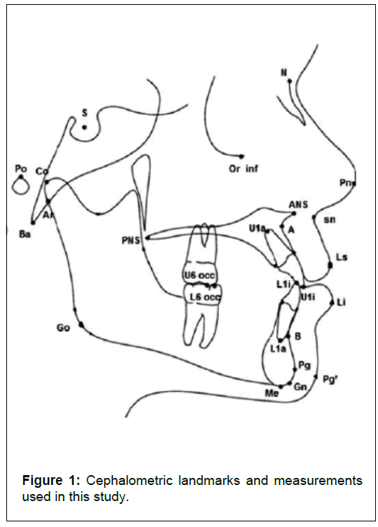
In group 1, electronic format of lateral cephalogram was imported in cephalometry software (dolphin imaging software version 11.95) and digitise tool was selected with the calibration scale set to 10 mm. Ruler points were placed on the image scale at 10 mm distance and landmarks for composite analysis were selected. The measure tool displayed measurements from which 10 linear and 10 angular values were chosen (Table 1).
| Angular values | |
|---|---|
| SNA | Angle determined by points S, N and A |
| SNB | Angle determined by points S, N and A |
| G Sn Pg | Angle formed between G, Sn and Pg |
| IMPA | Angle formed between Go-Me and the mandibular incisor axis |
| NLB | Angle determined by points columella, SN and UL |
| NbaPtGn | Angle formed between the Ba-N plane and the plane from Pt to Gn |
| Occ SN | Angle formed between occlusal plane and SN plane |
| SN FH | Angle formed between Frankfort horizontal (FH) and SN plane |
| U1 SN | Angle formed by the intersection of the maxillary incisor axis to SN plane |
| ANB | Angle formed between SNA and SNB angles |
| Linear values | |
| Chn Pg | Distance between hard and soft tissue pogonion |
| Co Gn | Distance between points Co and Gn |
| Co PtA | Distance from condylion to point A |
| LI NB | Perpendicular distance from the tip of the mandibular incisor to NB plane |
| NprPg | Distance between pogonion point and a line drawn perpendicular to FH from point N |
| NprPtA | Distance between point A and a line drawn perpendicular to FH from point N |
| U1 NA | Perpendicular distance from the tip of the maxillary incisor to NA plane |
| U6 PtV | Distance measured from distal surface of maxillary 1st molar to pteygoid vertical (PtV) |
| ULP | Distance measured from soft tissue point A to point A |
| Witts | Distance between points of A and B to the occlusal plane |
Table 1: The list of cephalometric measurements used for this study.
In group 2, radiographic film acquired from a DICOM printer was hand traced on acetate paper. Anatomical landmarks were identified and measurements for the chosen 20 parameters were done.
In group 3, the length and width of the radiographic image was measured using concepts (5.1, TopHatch, Inc), an iOS based app and was found to be 203.5 mm × 170.9 mm. This dimension was chosen as a standard for all radiographic image acquired from the same source. The dimensions were entered as a custom paper size setting in the consumer printer software with the option of fit to maximum size selected during print. Commercially available 180 gsm A4 size paper was chosen as per manufacturer recommendation for minimum quality printouts on a laser printer. The anatomical landmarks were identified and traced on acetate sheets attached to the printout (Figure 1).
The same parameters were followed as in the other two methods. Cephalometric analyses for all 10 digital lateral cephalograms were carried out using the three methods by a single operator and data was entered into excel.
Results
SPSS software for windows was used for the statistical analysis of the data. To test the statistical significance of the change in the values of the variables among the three methods, on an average, repeated measure analysis of variance was applied. In case of statistical significance, multiple comparison tests were applied to identify statistically significant pair of groups. P ≤ 0.05 was considered statistically significant.
There were no statistical difference in the computed values for 20 parameters except for one linear and three angular measurements. There was significant difference in the calculated mean between the digital (81.9 ± 4.9) and manual tracings (85.1 ± 4.3) for Co-PtA linear value. Similar statistical difference was found in the SNA, SNB and ANB angular measurements. The mean difference of SNA value was 83.4 ± 2.4 for digital, 81.5 ± 2.9 for manual and 81.8 ± 3.08 for paper print out; for SNB value was 80 ± 4.08 for digital, 78.9 ± 3.81 for manual and 78.9 ± 3.84 for paper print out and for ANB value was 2.1 ± 2.5 for digital, 3.7 ± 2.9 for manual and 3.3 ± 3.1 for paper print out (Tables 2 and 3).
| Digital (n=10) | Manual (n=10) | Paper (n=10) | ||||
|---|---|---|---|---|---|---|
| Mean | SD | Mean | SD | Mean | SD | |
| Linear values | ||||||
| Chn Pg' | 10.7 | 1.6 | 10.6 | 1.4 | 10.8 | 1.1 |
| L1 NB | 6.4 | 3.2 | 6.6 | 3.4 | 6.6 | 3.5 |
| ULP | 11.1 | 7.4 | 11.5 | 7.2 | 11.2 | 7.6 |
| Co Gn | 106.3 | 5.3 | 106.2 | 4 | 105.9 | 4.3 |
| NprPg | -7.2 | 5.9 | -6.3 | 6.7 | -6.8 | 7.3 |
| Witts | 0.7 | 4.7 | 0.3 | 3.3 | 0.1 | 3.6 |
| Co PtA | 81.9 | 4.9 | 85.1 | 4.5 | 84.8 | 5.7 |
| Npr PtA | -1.5 | 1.8 | -1.1 | 2.8 | -1.3 | 2.7 |
| U6 PtV | 17.1 | 5 | 16.7 | 6.9 | 16.3 | 6.7 |
| U1 NA | 5.9 | 2.5 | 6.3 | 2.4 | 6.1 | 2.1 |
| Angular values | ||||||
| G Sn Pg | 14 | 5.1 | 13.8 | 5 | 13.7 | 4.6 |
| IMPA | 99.1 | 9.1 | 101.1 | 13.7 | 101.6 | 14 |
| NLB | 97 | 34.9 | 98.1 | 14.9 | 98.6 | 14.4 |
| SN FH | 14.3 | 29.1 | 16.3 | 27.4 | 16.3 | 27.4 |
| Nba PtGn | 81.7 | 27.4 | 81 | 26.8 | 80.2 | 26.7 |
| Occ SN | 13 | 6.5 | 15 | 4.1 | 14.2 | 4.3 |
| SNA | 83.4 | 2.5 | 81.5 | 2.9 | 81.8 | 3.1 |
| SNB | 80 | 4.1 | 78.9 | 3.8 | 78.9 | 3.8 |
| U1 SN | 112.1 | 10 | 113.7 | 8.4 | 113.3 | 9.2 |
| ANB | 2.1 | 2.5 | 3.7 | 2.9 | 3.3 | 3.1 |
Table 2: Statistical evaluation of linear and angular values showing mean and standard deviation. Note: Linear and angular values referred to Table 1.
| Group 1 | Group 2 | Group 3 | F-test | 1_2 | 2_3 | 3_1 | |
|---|---|---|---|---|---|---|---|
| Angular measurements | |||||||
| SNA | 83.4+2.5 | 81.5+2.9 | 81.8+3.1 | * | * | * | - |
| SNB | 80.0+4.1 | 78.9+3.8 | 78.9+3.8 | * | * | * | - |
| G Sn Pg | 14.0+5.1 | 13.8+5.0 | 13.7+4.6 | ns | - | ||
| IMPA | 99.1+9.1 | 101.1+13.7 | 101.6+14.0 | ns | - | ||
| NLB | 97.0+34.9 | 98.1+14.9 | 98.6+14.4 | ns | - | ||
| NbaPtGn | 81.7+27.4 | 81.0+26.8 | 80.2+26.7 | ns | - | ||
| Occ SN | 13.0+6.5 | 15.0+4.1 | 14.2+4.3 | ns | - | ||
| SN FH | 14.3+29.1 | 16.3+27.4 | 16.3+27.4 | ns | - | ||
| U1 SN | 112.1+10.0 | 113.7+8.4 | 113.3+9.2 | ns | - | ||
| ANB | 2.1+2.5 | 3.7+2.9 | 3.3+3.1 | * | * | * | - |
| Linear measurements | |||||||
| Chn Pg | 10.7+1.6 | 10.6+1.4 | 10.8+1.1 | ns | - | ||
| Co Gn | 106.3+5.3 | 106.2+4.0 | 105.9+4.3 | ns | - | ||
| Co PtA | 81.9+4.9 | 85.1+4.5 | 84.8+5.7 | * | * | - | |
| LI NB | 6.4+3.2 | 6.6+3.4 | 6.6+3.5 | ns | - | ||
| NprPg | (-)7.2+5.9 | (-)6.3+6.7 | (-)6.8+7.3 | ns | - | ||
| NprPtA | (-)1.5+1.8 | (-)1.1+2.8 | (-)1.3+2.7 | ns | - | ||
| U1 NA | 5.9+2.5 | 6.3+2.4 | 6.1+2.1 | ns | - | ||
| U6 PtV | 17.1+5.0 | 16.7+6.9 | 16.3+6.7 | ns | - | ||
| ULP | 11.1+7.4 | 11.5+7.2 | 11.2+7.6 | ns | - | ||
| Witts | 0.7+4.7 | 0.3+3.3 | 0.1+3.6 | ns | - | ||
Note: *P<0.05; ns, not significant
Table 3: Statistical evaluation showing the significance of angular and linear values. Note: Linear and angular measurements referred to Table 1.
Discussion
In clinical orthodontics, cephalometric analysis has long been used as an important clinical tool in diagnosis, treatment planning and evaluation of growth or treatment results [7]. The major errors associated with conventional cephalometry include projection errors and tracing errors. The mechanical errors introduced by drawing lines between landmarks manually and by measuring with a ruler and protractor were common in conventional cephalometric analysis [8,9]. Because of the increasing use of cephalometric analysis for diagnosing malocclusion and treatment planning, the use of digital systems have risen in Orthodontics. The main advantages of digital radiology are the reduced radiation dose and improved data storage, information access and image manipulation. The most important criteria for using mechanical or digital method are that it should be accurate, precise and must show a high rate of reproducibility in both tracing and analysis. In recent time technology have contributed to production of affordable but high end consumer products. Such resources can be utilised when an alternative method becomes necessary. Hence the prime objective of this study was to compare the accuracy of lateral cephalograms traced manually, digitally and on paper printout. The present study evaluated the reliability of cephalometric measurements obtained using a computerized program on direct digital radiographs as well as with the handtracing method. Identification of the landmarks is important as the tracing method because interoperator error is found to be greater than intraoperator error [10]. As a result the measurements were carried by one examiner only.
Since there was no gold standard method for calibration of digital radiograph image all three methods were compared. The results of the analyses showed that the measurements performed were independent of the type of tracing done (digital, manual or paper). The parameters used in this study were commonly used cephalometric measurements for assessing different kinds of malocclusion in orthodontic diagnosis and treatment planning which included 10 linear and 10 angular measurements.
In the present study, there were no statistical significant differences except in one linear (CoPtA) and three angular measurements (SNA, SNB and ANB) (p ≤ 0.05). The reason behind the statistical significance in the three methods may be due to calibration error or difference in landmark identification. The three angular values which showed statistically significant differences were SNA, SNB and ANB. The parameters comprised of locating common landmarks, Sella (S) and Nasion (N). Statistical differences were found in manual tracing compared with digital software and paper tracing, whereas no difference was present between the software and paper tracing.
Measurements obtained from digital tracing and manual tracing were shown to have adequate reproducibility [11]. These findings coincide with the present study result. However the digital and manual tracing cephalometry are compared which gave a statistically significant differences between measurements which are not in accordance with our study results [12].
Both methods of conventional and digital cephalometric analysis are highly reliable with some statistically significant differences in reproducibility but most were not clinically significant [13]. It provides support for computerized tracing method as these are easier and less time consuming with same reliability when compared to manual tracing [14]. The reliability of cephalometric measurements on the digital cephalogram is generally comparable to those on original radiographs [15]. In order to obtain a quantitative and objective evaluation of the accuracy of cephalometric measurements, a large sample size is essential. This was the disadvantage of the present study.
Conclusion
DICOM printout showed significant difference in three angular and one linear values of the total 20 values when compared with other methods. Consumer paper printout tracing is a reliable option when printer settings are customised to provide 1:1 prints.
References
- Brodie AG (1941) On the growth pattern of the human head from the third month to the eighth year of life. Am J Anat 68: 209-262.
- Baumrind S, Frantz RC (1971) The reliability of head film measurements: 2 Conventional angular and linear measures. Am J Orthod 60: 505-517.
- Ricketts RM (1981) Perspectives in the clinical application of cephalometrics: The first fifty years. Angle Orthod 51: 115-150.
- Sandler PJ (1988) Reproducibility of cephalometric measurements. Br J Orthod 15: 105-110.
- Carlos Quintero J, Trosien A, Hatcher D, Kapila S (1999) Craniofacial imaging in orthodontics: Historical perspective, current status and future developments. Angle Orthod 69: 491-506.
- Brannan J (2002) An introduction to digital radiography in dentistry. J Orthod 29: 66-69.
- Chen YJ, Chen SK, Chung-Chen Yao J, Chang HF (2004) The effects of differences in landmark identification on the cephalometric measurements in traditional versus digitized cephalometry. Angle Orthod 74: 155-161.
- Gravely JF, Benzies PM (1974) The clinical significance of tracing error in cephalometry. Br J Orthod 1: 95-101.
- Cohen AM (1984) Uncertainty in cephalometrics. Br J Orthod 11: 44-48.
- Sayinsu K, Isik F, Trakyali G, Arun T (2007) An evaluation of the errors in cephalometric measurements on scanned cephalometric images and conventional tracings. Eur J Orthod 29: 105-108.
- Grybauskas S, Balciuniene I, Vetra J (2007) Validity and reproducibility of cephalometric measurements obtained from digital photographs of analogue headfilms. Stomatologija Baltic Dental and Maxillofacial J 9: 114-120.
- Collins J, Shah A, McCarthy C, Sandler J (2007) Comparison of measurements from photographed lateral cephalograms and scanned cephalograms. Am J Orthod Dento Ortho 132: 830-833.
- Albarakati SF, Kula KS, Ghoneima AA (2012) The reliability and reproducibility of cephalometric measurements: A comparison of conventional and digital methods. Dentomaxil Radiol 41: 11-17.
- Prabhakar R, Rajakumar P, Karthikeyan MK, Saravanan R, Vikram R, et al. (2014) A hard tissue cephalometric comparative study between hand tracing and computerized tracing. J Pharm Bioal Sci 6: S101.
- Chen SK, Chen YJ, Yao CCJ, Chang HF (2004) Enhanced speed and precision of measurement in a computer-assisted digital cephalometric analysis system. Angle Orthod 74: 501-507.
Open Access Journals
- Aquaculture & Veterinary Science
- Chemistry & Chemical Sciences
- Clinical Sciences
- Engineering
- General Science
- Genetics & Molecular Biology
- Health Care & Nursing
- Immunology & Microbiology
- Materials Science
- Mathematics & Physics
- Medical Sciences
- Neurology & Psychiatry
- Oncology & Cancer Science
- Pharmaceutical Sciences

