Simple And Chairside Construction And Placement Of Guide For Accurate Positioning Of Orthodontic Mini- Implants
Dr. Vaibhav Gandhi and Dr. Falguni Mehta
Dr. Vaibhav Gandhi* Dr. Falguni Mehta
Department of orthodontics, Govt. Dental College and Hospital, Ahmedabad-380009, India
- *Corresponding Author:
- Dr. Vaibhav Gandhi
3rd Year Post Graduate, Department of orthodontics
Govt. Dental College and Hospital
Ahmedabad-380052,India
Tel: +919898562563
E-mail: smiledesigner1988@gmail.com
Introduction
Conservation of anchorage in totality has been a perennial problem for all the orthodontists. Conventional means of supporting anchorage use either intra-oral sites or rely on extra oral means. Both of these have their own limitations. The extra-oral forces cannot be used on a 24 hours a day to resist the continuous tooth moving forces and are dependent on the patient's compliance. Intraoral conventional methods also encounter some percentage of anchor loss, although patient compliance is not an issue. Studies of Prof. Branemark, a pioneer in osseointegrated implants have opened new horizon in field of dentistry. Micro implants were introduced for orthodontic usage. These orthodontic implants made from titanium have changed the traditional concepts for anchorage. They are now widely used as “Absolute Anchorage” which is not dependent on dental units for its anchorage [1].
But there are chances that some complications can be arise during treating case with implant anchorage like mobility of micro implants, oro-antral communication, peri-implantitis, proximity of tooth root, undesirable tooth movement and micro implant fracture. The proximity of micro implants to the adjacent tooth root is a major risk factor for failure of implants. This is more common in the mandible, suggesting that micro implants’ placement needs to be accurate to avoid root proximity and loosening of implants. Micro implant when touching the root may cause changes from root resorption to loss of vitality, osteosclerosis and dentoalveolar ankylosis. Hence precise placement of implant is necessary for its success. “Guides” help in precise placement of implants and are therefore necessary for accurate placement of implants thereby avoiding such complications [2,3].
Method of construction Guide
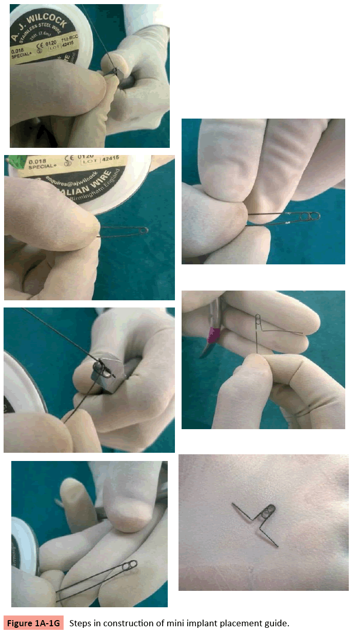
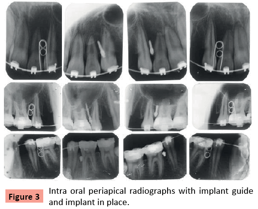
Simple basic instruments like 0.018’’ A.J.Wilcock stainless steel wire, 139 plier and glass marking pencil are required. Steps for making double helical implant guide is shown in (Figure 1A-1G). Photographs with guide placed in position is shown in (Figure 2). To check the final position of implants, IOPAs (intraoral periapical radiographs) are required. (Figure 3) Bisecting technique was used to take radiograph.
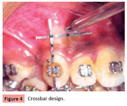
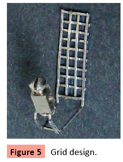
There is a list of advantages of this implant placement guide like minimum armamentarium required, chairside construction with easy and simple wire bending, easy to place with lig-a-ring on the bracket, remains stable for accurate implant placement, IOPA can be easily taken after guide placement, no need to remove base arch wire for guide placement, implants can be placed with guide already in place, easy Placement and removal, comfortable to the patient and can be made for any site. Other designs like crossbar requires welding machine as well as base arch wire must have to be removed, which are not required in our design [4] and Grid design is too complex design to make as well as chair side construction is not possible [5].
References
- Rajesh P, Girish K, Manish S (2011) Orthodontic Microimplants and Its Application. Journal of Contemporary Dentistry Jun-Sept 1,1
- Kuroda S, Yamada K, Deguchi T, Hashimoto T, Kyung HM, Takano-Yamamoto T (2007) Root proximity is a major factor for screw failure in orthodontic anchorage. Am J Orthod Dentofacial Orthop; 131:00
- Farinazzo Vitral RW, Santiago RC, Oliveira GS, Fraga MR, da Silva Campos MJ (2012) Mini-Implants: When Orthodontists are caught in their Own Web. J Clin Case Rep 2:130.
- Belludi et al: Crossbar: An effective orthodontic mini implant placement Guide: J Of Ind Ortho Soc 2010; 44: 149-151
- Bharanikumar R, Pavankumar .M, Nagendrakumar.M (2008) A grid for guiding miniscrew placement. J.Clin.orthod. :531-532
Open Access Journals
- Aquaculture & Veterinary Science
- Chemistry & Chemical Sciences
- Clinical Sciences
- Engineering
- General Science
- Genetics & Molecular Biology
- Health Care & Nursing
- Immunology & Microbiology
- Materials Science
- Mathematics & Physics
- Medical Sciences
- Neurology & Psychiatry
- Oncology & Cancer Science
- Pharmaceutical Sciences
