Treatment of a Class II Division 2 Malocclusion in a Teenage Patient: Clinical Case Report
Ramírez Silvia,Siguencia Valeria,García Andrés and Bravo Manuel
DOI10.21767/2469-2980.100047
Ramírez Silvia1*, Siguencia Valeria2, García Andrés3 and Bravo Manuel4
1Dentist, Resident student of the Orthodontic Specialization of the University of Cuenca
2Specialist in Orthodontics and Maxillofacial Orthopedics. Professor of Orthodontics at the University of Cuenca. Member of the Ecuadorian Society of Orthodontics
3Specialist in Orthodontics and Maxillofacial Orthopedics. Professor of Orthodontics at the University of Cuenca
4Doctor of Dentistry, University of Cuenca. Master in Orthodontics, C. University of Sao Paulo-Brazil 2010. Member of the World Federation of Orthodontics. Member of the American Association of Orthodontics. Member of the Ibero-American Society of Lingual Orthodontics. Member of the Spanish Society of Orthodontics. Member of the Ecuadorian Society of Orthodontics. Member of the Society of Orthodontics and Orthopedics of Pichincha. Member of the Society of Orthodontics and Orthopedics of Azuay
- *Corresponding Author:
- Silvia R
Dentist, Resident Student of the Orthodontic Specialization
Universidad de Cuenca
Cuenca, 010162 Ecuador
Tel: +593998444452
E-mail: silvia.ramirez@ucuenca.ec
Received Date: October 20, 2017; Accepted Date: November 14, 2017; Published Date: November 21, 2017
Citation: Silvia R, Valeria S, Andrés G, Manuel B (2017) Adolescent patient; Treatment; Maxillary molars; Space; Crowding. J Orthod Endod 3:13. doi: 10.21767/2469-2980.100047
Abstract
This article describes the orthodontic treatment of an adolescent patient presenting a class II skeletal, convex profile, mesofacial biotype, upper dental midline deviated 1 mm to the left, Class I bilateral molar, canine distoclusion of ½ right unit and left canine relationship non-determinable because piece 23 is in ectopic position, proinclination and inferior protrusion. The treatment plan was to distalize the maxillary molars and create enough space to incorporate pieces 13, 23 in the dental arch, a pendulum appliance supported with two orthodontic mini implants were used. The active treatment lasted 18 months and at the end of it, all the objectives were fulfilled, resulting in facial balance. The pendulum appliance is a good alternative for a Class II dental correction, it produces distalization of the maxillary molars in an optimal treatment time.
Keywords
Adolescent patient; Treatment; Maxillary molars; Space; Crowding
Introduction
Class II malocclusions can be corrected in various ways, such as reorientation of maxillary growth, distal movement of the maxillary dentition, mesial movement of the mandibular dentition and increase or reorientation of mandibular growth [1]. When the treatment plan for the patient is considered, the question arises: Do we need to remove the teeth or create necessary space without extractions? Before this question, when it is chosen to create the necessary space without extractions, the professional must find alternatives to obtain the necessary space to correct the malocclusion. A considerable decrease in the percentage of extractions as part of the orthodontic treatment has been observed in recent years, since with these extractions, stability at the end of the treatment is not necessarily obtained. Molar distalization is a treatment option that aims to create additional space within the arch, being of great value in cases where there is a normal mandibular relationship, minimal bonetooth discrepancy, Class II molar relationship, and it is desired to avoid extractions of permanent teeth, in the end one obtains a Class I molar relationship and the necessary space in the lateral segments of the canines or premolars. In this way, anterosuperior crowding is resolved by moving the molars to distal in the initial stages of treatment [2-4].
Nowadays, there is a great variety of devices for the distalization of upper molars, not long ago "extraoral traction" was one of the devices most used to distalize; however, due to its disadvantages such as the need for cooperation on the part of the patient and very little acceptance, it has been replaced by a series of intraoral devices such as Niti springs, Sliding Jig, Distal jet, Dual Force distalizer, Cortical Dual Force Distalizer among others. An intraoral system to move molars distally was introduced in 1992 by Hilgers [5-7].
This case report describes the distalization of upper molars obtaining the necessary space for the correct placement of pieces 13, 23 in the dental arch, using a pendulum appliance. Among its advantages it has been described: Minimum need of collaboration from the patient; acceptable aesthetics and comfort; minimum loss of anterior anchorage; body movement of the molars to avoid undesirable side effects such as lengthening of treatment and unstable results; minimum time for placement and reactivation [8].
Diagnosis
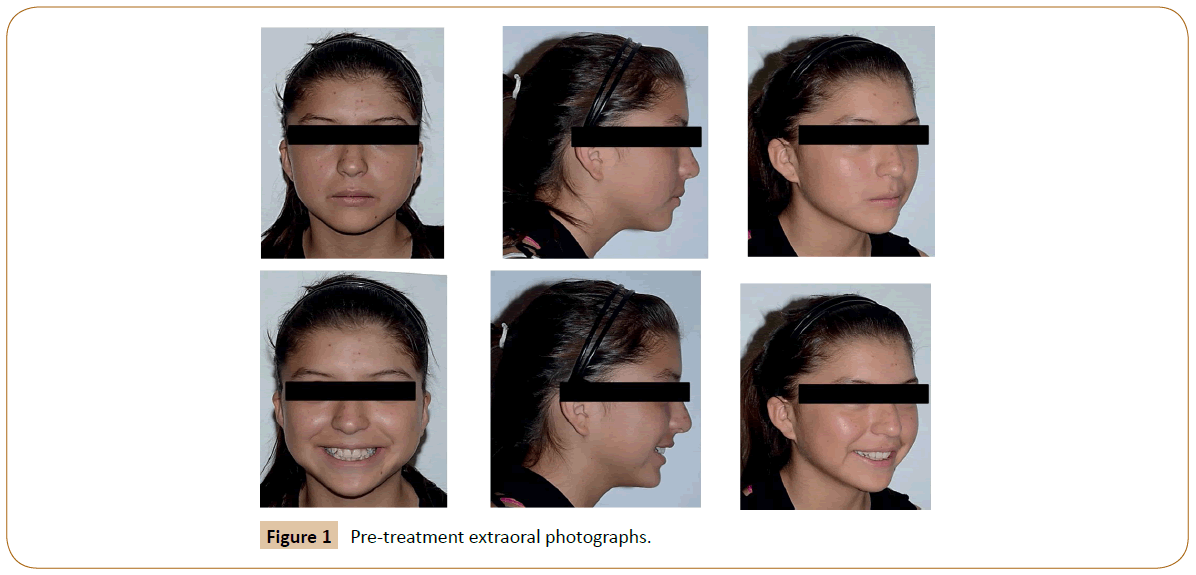
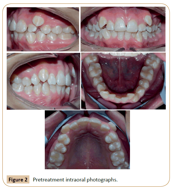
The patient is a female, age 12 years 5 months, presented a class II skeletal, convex profile, mesofacial biotype, mandibular rotation straigh down, superior dental midline deviated 1 mm to the left, bilateral molar neutroclusion, canine distoclusion of ½ right unit and an indeterminable left canine distoclusion relationship, Spees curve of -2.5 mm, overjet of 3 and 3 mm overbite, proinclination and protrusion inferior, total superior discrepancy of -11.4 total inferior discrepancy of -8.4 (Figures 1 and 2).
Objectives of the Treatment
The objectives of the treatment were:
1. To achieve bilateral canine neutroclusion and class I bilateral molar.
2. Level Spees curve.
3. Incorporate parts 13 and 23 into the dental arch.
4. Overbite and protrusion correction.
5. Correct and align all incorrect positions.
6. Correct upper and lower dental midline.
Alternatives of Treatment
Among the treatment alternatives, we considered:
1. Non-conservative treatment: Surgical extraction of the first four premolars due to the superior and inferior tooth bone discrepancy. Upper and lower fixed appliance prescription MBT slot 0.022.
2. Non-conservative treatment: Surgical extraction of the first two upper premolars because the greatest tooth bone discrepancy is at the level of the upper arch. Upper and lower fixed appliance prescription MBT slot 0.022.
3. Conservative treatment: Distalization of maxillary molars by pendulum with bone anchorage, with the aim of obtaining space for the canines and achieving the relation of bilateral neutroclusion. Upper and lower fixed appliance prescription MBT slot 0.022.
The patient did not accept the extractions of the first premolars and distalization of the maxillary molars proceeded.
Treatment Progress
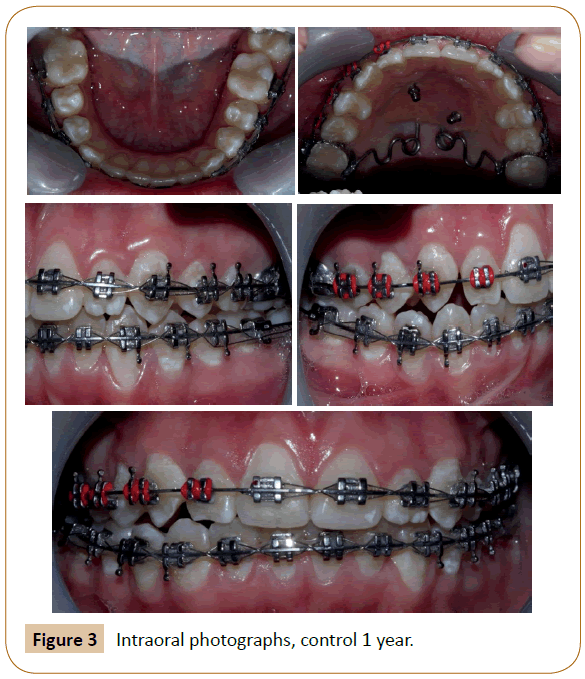
The impression of the upper arch is used to create the pendulum. Once ready, it was placed in the patient's mouth and anchored with two miniscrew (2.0 mm diameter 10.0 mm length); before being cemented, the activation of each arm was performed at 45º. We made the assembly of the fixed appliance prescribed MBT slot 0.022 in both the upper arch and the lower arch, all pieces were included in the arch except for pieces 13, 23 due to lack of space. An initial upper and lower Niti arc 0.012 was used for the existing crowding, and then was changed to an upper and lower Niti arc 0.014, as pieces 16, 26 were distalized, the anterosuperior crowding was gradually corrected. The sequence of upper and lower Niti arches 0.016; 0.016 x 0.01; 0.016 x 0.022 was continued. After six months of treatment, sufficient space could be observed for the inclusion of pieces 13, 23. The same pendulum was used as containment for six more months of the treatment, working with class II Vector 3/16" 41/2 oz. elastic of the right side and triangular ligaments started to maintain neutroclusion relation of the left side (Figure 3).
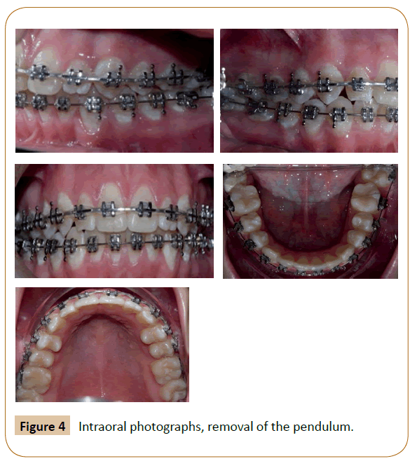
One year after treatment, the pendulum was removed and work continued with the alignment, leveling and final details of the treatment (Figure 4).
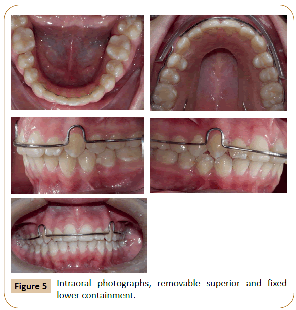
At one and a half years, all the objectives of the treatment were obtained, the appliance was removed, fixed lower multistrand bonded retainer and an upper hawley type retainer with continuous vestibular arch were indicated (Figure 5).
Treatment Results
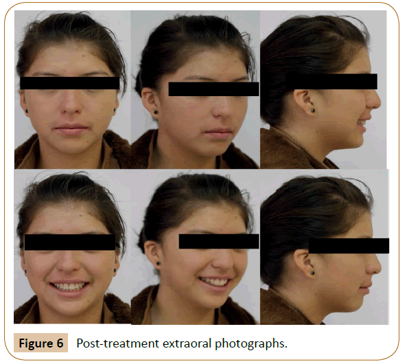
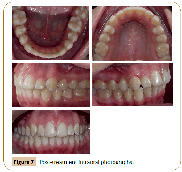
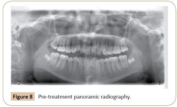
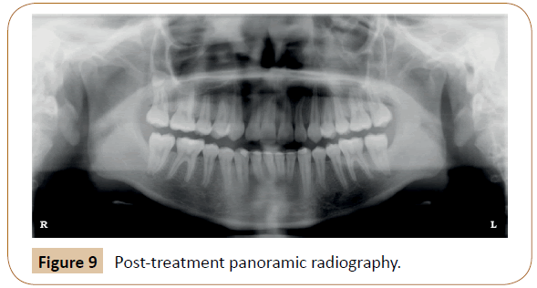
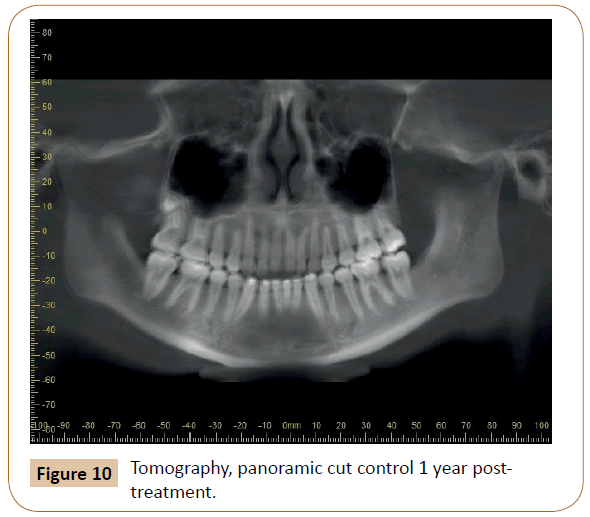
All the proposed objectives were fulfilled: A class I bilateral angle molar, bilateral canine neutroclusion relation was achieved, crowding was eliminated, upper and lower dental midline coincidence was obtained, adequate Overjet and Overbite, with the patient ending up with a harmonious smile. Regarding cephalometric measures, changes were observed (Figures 6-8).
Discussion
"Extraction versus non-extraction" is a subject that has been widely discussed in orthodontic literature. Before the "NO EXTRACTION," the option of "distalization" has arisen, which is preferred when the patient has an acceptable profile accompanied by a bad class II molar occlusion half cusp or less. Sometimes distalization is the only viable option, because the patient presents in the dental office with previous orthodontic treatment, and having had upper premolar extraction and excessive overjet [9].
Multiple studies have shown that care should be taken with the use of extraoral devices for distalization of molars in which patient collaboration is required, as there is a distal molar movement accompanied by a loss of anchorage that is evidenced in the mesial movement of the incisors and premolars. Therefore, it is necessary to consider several treatment alternatives [10].
Intraoral distalization appliances have been designed, which generate a continuous reciprocal force in the first upper molars. Initially, all the cited appliances succeeded in distalizing the molars successfully in the upper maxilla, despite the space obtained between the first molar and second premolar or a primary molar, this success must also be based on the reaction of the anchor unit. The anchor loss causes a mesialization effect on the anchor components. Consequently, if the premolars or incisors (or both) are the anchoring teeth, they move mesially, the incisors protrude, and the overjet increases [11,12].
Among the intraoral distalizers are the dento-supported, among which the best known is the Hilgers Pendulum [5,6,12]. The pendulum has undergone widespread clinical use, several studies have demonstrated its skeletal and dentoalveolar effects [3,6,11]. It was found that the pendulum is an effective device in the distalization of maxillary molars, but it is also accompanied by a loss of anterior anchorage as cited [11,13], this being approximately 30 to 43% of the space created between the molars and premolars because of their dental anchorage.
In recent years, thanks to the appearance of mini-implants and mini-plates, osteo-dental supported distalizers have emerged, which aim to counteract these side effects [5,14]. In their design, they present a bony anchorage in the anterior part. When using mini-implants, intraoral anchoring can be achieved without incorporating teeth [15].
Molar distalization when using the pendulum with both anchoring systems is effective. The distal molar movement recorded was 3.34 mm with conventional anchorage and 5.10 mm with the skeletal anchorage system. The conventional anchoring system showed mesial premolar movement of 4.01 mm, unlike the skeletal anchorage system which did not show anchorage loss; there was a spontaneous distal movement of 2.30 mm in the premolar when direct anchorage was used. Therefore, intraoral distalizers associated with direct skeletal anchorage are a viable method to minimize the effects of anchorage loss in the treatment of Class II malocclusions [16].
In a systematic review by Fontana et al., it was found that in patients with skeletal anchorage system, the movement varied from 6.4 mm to 0.5 mm and a distal inclination of 18.5°. Loss of premolar anchoring and incisor proclination are an unavoidable side effect ranging from 4.33 mm to 0.73 mm and 13.7° to 0.6° respectively. Vertical skeletal changes were also observed, which was evidenced in an increase in the vertical facial dimension varying from 1.5° to -1.8° [17]. The results indicate that there is a predominantly dental action, with no significant alterations in the vertical dimension Table 1 [3,18,19].
Byloff et al. concluded that the pendulum appliance produces 3.39 mm ± 1.25 mm of distal movement with a mean bimolar intrusion of 1.17 mm ± 1.29 mm [14]. With the distalization of the upper molar, 76% of the total opening of the anterior space to the maxillary first molar was obtained. The upper central incisors bent slightly during treatment [20].
In this case report, a distalization of 5.9 mm was obtained and at the level of the upper incisors a protrusion of 3.4 mm was produced, accompanied by a proclination of 2° (Figures 8-10), that is to say the space was obtained thanks to the molar distalization but also due to the secondary effect of the incisal protrusion; it was evidenced that the distalization with pendulum skeletal anchorage is accompanied by the secondary effect of incisal protrusion, results that are supported in the majority of performed revisions.
According to Kizinger et al., the right moment to initiate distalization with a pendulum is before the eruption of the second molars. There are few studies that describe distalization in the presence of the fully erupted second molar; if the former is to be performed it must be done in stages, first the second molars and then the first molars, once a germectomy of the third molar has been performed as a first procedure. It has been seen that there is a greater loss of anchorage and recurrence of the second molar. The patient in our report had the second molars fully erupted, in occlusion; nevertheless at six months of beginning the treatment the space necessary for the correct alignment of all the pieces in the arch was obtained [21-23].
For Kizinger et al., molar distalization and protrusion of the incisors are directly correlated with the patient's dental age, they occur in a greater proportion in the early mixed dentition than in the permanent dentition, with the protrusion of the incisors being less pronounced. Without a doubt, the age of the patient greatly influenced the moment of the distalization, allowing us to achieve the desired space in an optimal time of treatment. Orthodontic treatment simultaneously with distalization of the maxillary molars shortened the total treatment time [24,25].
Conclusion
This case report shows that the pendulum with skeletal anchorage is a good alternative for dental Class II correction by distalizing the maxillary molars in an optimal treatment time. All the objectives were met and the patient at the end of the treatment obtained a smile that complied with the parameters of micro aesthetics and macro aesthetics, a suitable occlusion with canine disocclusion, anterior guide and a harmonious profile.
Conflict of Interest
None.
References
- Lalitha Ch, Vasumurthy S, Vikasini K (2011) Recent advances of Pendulum appliance for effective molar distalization. Indian J Dent Adv 3: 572-576.
- Vasanthan P, Sabarinathan J, Sabitha S, Sathishkumar RC (2015) Reliable molar distalizer: A review. J Adv Clin Res Insights 2: 40-43.
- Kinzinger GSM, Diedrich PR (2007) Biomechanics of a modified Pendulum appliance-theoretical considerations and in vitro analysis of the force systems. Eur J Orthod 29: 1-7.
- Ciro P, Sandoval P, Rey D, Uribe G, Sierra A, et al. (2011) Distalización de molares maxilares con aparatos intraorales de nueva generación que no necesitan colaboración del paciente. Int J Odontostomat 5: 39-47.
- Campuzano A, Siegert M, Rey D (2014) Distalización con el C-DFD modificado con mini-tornillos. Reporte de caso. CES Odontología 27: 131-141.
- Hilgers JJ (1992) The Pendulum appliance for Class II non-compliance therapy. J Clin Orthod 26: 706-714.
- Taner TU, Yukay F, Pehlivanoglu M, Cakirer B (2003) A comparative analysis of maxillary tooth movement produced by cervical headgear and pend-x appliance. Angle Orthod 73: 686-691.
- Chen G, Teng F, Xu TM (2016) Distalization of the maxillary and mandibular dentitions with miniscrew anchorage in a patient with moderate Class I bimaxillary dentoalveolar protrusion. Am J Orthod Dentofacial Orthop 149: 401-410.
- Noorollahian S, Alavi S, Shirban F (2015) Non-compliance appliances for upper molar distalization: An overview. Int J Orthod Milwaukee 26: 31-36.
- Antonarakis GS, Kiliaridis S (2008) Maxillary molar distalization with noncompliance intramaxillary appliances in Class II malocclusion. A systematic review. Angle Orthod 78: 1133-1140.
- Kircelli BH, PektaÃÆââ¬Â¦Ãâà ¸ ZO, Kircelli C (2006) Maxillary molar distalization with a bone-anchored pendulum appliance. Angle Orthod 76: 650-659.
- Kinzinger GSM, Eren M, Diedrich PR (2008) Treatment effects of intraoral appliances with conventional anchorage designs for non-compliance maxillary molar distalization: A literature review. Eur J Orthod 30: 558-571.
- Sugawara J, Kanzaki R, Takahashi I, Nagasaka H, Nanda R (2006) Distal movement of maxillary molars in nongrowing patients with the skeletal anchorage system. Am J Orthod Dentofac Orthop. 129: 723-733.
- Byloff FK, Darendeliler MA (1997) Distal molar movement using the pendulum appliance. Part 1: Clinical and radiological evaluation. Angle Orthod 67: 249-260.
- Kinzinger GSM, Gross U, Fritz UB, Diedrich PR (2005) Anchorage quality of deciduous molars versus premolars for molar distalization with a pendulum appliance. Am J Orthod Dentofac Orthop 127: 314-323.
- Grec RH, Janson G, Branco NC, Moura-Grec PG, Patel MP, et al. (2013) Intraoral distalizer effects with conventional and skeletal anchorage: a meta-analysis. Am J Orthod Dentofac Orthop 143: 602-615.
- Fontana M, Cozzani M, Caprioglio A (2012) Non-compliance maxillary molar distalizing appliances: An overview of the last decade. Prog Orthod 13: 173-184.
- Chaqués-Asensi J, Kalra V (2001) Effects of the pendulum appliance on the dentofacial complex. J Clin Orthod 35: 254-257.
- Silva FLGR, Barbosa HAM, Oliveira DTN, Bertoz APM, Faltin Jr. K, et al. (2016) Dental changes in Class II patients treated with Pendex appliance: A prospective study. Arch Health Investig. 5: 186-191.
- Bussick TJ, McNamara JA (2000) Dentoalveolar and skeletal changes associated with the pendulum appliance. Am J Orthod Dentofac Orthop 117: 333-343.
- Kinzinger GSM, Fritz UB, Sander FG, Diedrich PR (2004) Efficiency of a Pendulum appliance for molar distalization related to second and third molar eruption stage. Am J Orthod Dentofac Orthop 125: 8-23.
- Kinzinger GSM, Wehrbein H, Gross U, Diedrich PR (2006) Molar distalization with Pendulum appliances in the mixed dentition: Effects on the position of unerupted canines and premolars. Am J Orthod Dentofac Orthop 129: 407-417.
- Caprioglio A, Cozzani M, Fontana M (2014) Comparative evaluation of molar distalization therapy with erupted second molar: Segmented versus Quad Pendulum appliance. Prog Orthod 15: 49.
- Kinzinger GSM, Wehrbein H, Diedrich PR (2005) Molar distalization with a modified pendulum appliance-in vitro analysis of the force systems and in vivo study in children and adolescents. Angle Orthod 75: 558-567.
- Chiu PP, McNamara JA, Franchi L (2005) A comparison of two intraoral molar distalization appliances: Distal jet versus pendulum. Am J Orthod Dentofac Orthop 128: 353-365.
Open Access Journals
- Aquaculture & Veterinary Science
- Chemistry & Chemical Sciences
- Clinical Sciences
- Engineering
- General Science
- Genetics & Molecular Biology
- Health Care & Nursing
- Immunology & Microbiology
- Materials Science
- Mathematics & Physics
- Medical Sciences
- Neurology & Psychiatry
- Oncology & Cancer Science
- Pharmaceutical Sciences
